

Diseases of the corneal endothelium
- PMID: 33596440
- PMCID: PMC8044020
- DOI: 10.1016/j.exer.2021.108495
Diseases of the corneal endothelium
Abstract
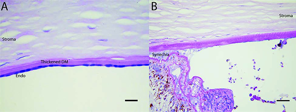
The corneal endothelial monolayer and associated Descemet's membrane (DM) complex is a unique structure that plays an essential role in corneal function. Endothelial cells are neural crest derived cells that rest on a special extracellular matrix and play a major role in maintaining stromal hydration within a narrow physiologic range necessary for clear vision. A number of diseases affect the endothelial cells and DM complex and can impair corneal function and vision. This review addresses different human corneal endothelial diseases characterized by loss of endothelial function including: Fuchs endothelial corneal dystrophy (FECD), posterior polymorphous corneal dystrophy (PPCD), congenital hereditary endothelial dystrophy (CHED), bullous keratopathy, iridocorneal endothelial (ICE) syndrome, post-traumatic fibrous downgrowth, glaucoma and diabetes mellitus.
Keywords: Edema; Endothelial disease; Fibrosis; Fuchs corneal endothelial dystrophy; Stroma.
Copyright © 2021 Elsevier Ltd. All rights reserved.
Figures

References
-
- Adamis AP, Filatov V, Tripathi BJ, Tripathi R.A.m.C., 1993. Fuchs’ endothelial dystrophy of the cornea. Survey of Ophthalmology 38, 149–168. - PubMed
-
- Aldave AJ, Yellore VS, Yu F, Bourla N, Sonmez B, Salem AK, Rayner SA, Sampat KM, Krafchak CM, Richards JE, 2007. Posterior polymorphous corneal dystrophy is associated with TCF8 gene mutations and abdominal hernia. American Journal of Medical Genetics Part A 143A, 2549–2556. - PubMed
Publication types
MeSH terms
Supplementary concepts
Grants and funding
LinkOut - more resources
Full Text Sources
Other Literature Sources
Medical
