. 2021 May 12;121(9):5597-5631.
doi: 10.1021/acs.chemrev.0c01137. Epub 2021 Feb 17.

The Whole Is Bigger than the Sum of Its Parts: Drug Transport in the Context of Two Membranes with Active Efflux

Affiliations

The Whole Is Bigger than the Sum of Its Parts: Drug Transport in the Context of Two Membranes with Active Efflux

Valentin V Rybenkov et al. Chem Rev. .

Abstract

Cell envelope plays a dual role in the life of bacteria by simultaneously protecting it from a hostile environment and facilitating access to beneficial molecules. At the heart of this ability lie the restrictive properties of the cellular membrane augmented by efflux transporters, which preclude intracellular penetration of most molecules except with the help of specialized uptake mediators. Recently, kinetic properties of the cell envelope came into focus driven on one hand by the urgent need in new antibiotics and, on the other hand, by experimental and theoretical advances in studies of transmembrane transport. A notable result from these studies is the development of a kinetic formalism that integrates the Michaelis-Menten behavior of individual transporters with transmembrane diffusion and offers a quantitative basis for the analysis of intracellular penetration of bioactive compounds. This review surveys key experimental and computational approaches to the investigation of transport by individual translocators and in whole cells, summarizes key findings from these studies and outlines implications for antibiotic discovery. Special emphasis is placed on Gram-negative bacteria, whose envelope contains two separate membranes. This feature sets these organisms apart from Gram-positive bacteria and eukaryotic cells by providing them with full benefits of the synergy between slow transmembrane diffusion and active efflux.


Conflict of interest statement

The authors declare no competing financial interest.

Figures

Figure 1.
Figure 1.
Organization of the cell envelope in Gram-negative bacteria on the example of E. coli. Key components of the cell envelope that define penetration of small molecules into bacteria include the inner and outer membranes, porins (such as OmpF), efflux transporters that act across the outer membrane (such as AcrAB-TolC), and substrate specific importers and exporters such as LacY and MdfA, respectively. The crystal structures shown are OmpF (PDB 2ZLD), LacY (PDB 2V8N), MdfA (PDB 4ZOW), and AcrAB-TolC (PDB 5O66).
Figure 2.
Figure 2.
Contrasting partition (P) and distribution (D) coefficients. Uncharged species of weak acids and bases dominate transmembrane permeation and partitioning into organic phase. HA and HAM are, respectively, the water- and membrane-soluble fractions of the protonated form of the acid.
Figure 3.
Figure 3.
Measured and predicted permeabilities of various antibiotics. Experimental data were collected from refs – and represent measurements using artificial lipid bilayers (lipids) or whole cells, as indicated. The predicted permeabilities were calculated using eq 1b with octanol–water clogD7.4 values computed at the Collaborative Drug Discovery Database (Burlingame, CA, www.collaborativedrug.com), assuming the thickness of the bilayer of 3 nm and the diffusion coefficient in hexadecane that was corrected for the size of the solvent molecules but not the shape of the solute.
Figure 4.
Figure 4.
A multilayer slab model of a membrane. Properties in each slab mimic those of the bilayer.
Figure 5.
Figure 5.
Lipid assemblies: (A) micelles, (B) unilamellar liposomes, (C) multilamellar liposomes, (D) supported mono- and bilayers, (E) immobilized artificial membranes, (F) planar bilayers (black lipids), (G) parallel artificial membrane permeability assay, and (H) Caco-2 permeability assay.
Figure 6.
Figure 6.
Crystal structure of OmpF, PDB 1OPF (A), and LamB, PDB 1MAL (B). Key residues involved in substrate binding are shown as sticks including residues D113, E117, R42, R82, and R132 on OmpF and residues W74, Y41, Y6, W420, W358, F227, and Y118 of LamB.
Figure 7.
Figure 7.
Three steps of ampicillin translocation through OmpF as predicted using molecular dynamics simulations. The residues involved in hydrogen bonding with ampicillin are shown in ball-and-stick representation (basic, blue; acidic, red; hydrophobic, gray), and the hydrophobic pockets are shown as a gray surface. Reprinted with permission from ref . Copyright 2010 American Chemical Society.
Figure 8.
Figure 8.
Electrophysiology analysis of porins reconstituted in planar lipid bilayers.
Figure 9.
Figure 9.
Representative examples of ABC uptake and efflux transporters of E. coli acting in the context of two membranes and their kinetic properties. The crystal structures shown are: LamB (PDB 1MPQ), MalE (PDB 2FNC), MalEFGK (PDB 2R6G), and MacAB-TolC (PDB 5NIK).
Figure 10.
Figure 10.
A genetic approach for controlled variation of the OM permeability and active efflux. A set of isogenic strains that differ in the expression of select efflux pumps and a nonspecific OM pore is used for the side-by-side quantification of intracellular accumulation of compounds.
Figure 11.
Figure 11.
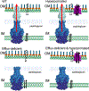
Kinetic mechanism of small molecule penetration across the Gram-negative cell envelope.
Figure 12.
Figure 12.
Steady-state accumulation levels of small molecules that cross a membrane with an efflux transporter (A), two efflux transporters (B), or an uptake transporter (C). The solution to the system of one efflux transporter (A) is given by R function (eq 19). α is a numeric constant traceable to B that varies from one system to another.
Figure 13.
Figure 13.
Increase in the MIC due to active efflux. Adapted with permission from ref . Copyright 2020 American Chemical Society.

References

    1. Nikaido H Prevention of Drug Access to Bacterial Targets: Permeability Barriers and Active Efflux. Science 1994, 264, 382–388. - PubMed
    1. Zgurskaya HI; Rybenkov VV Permeability Barriers of GramNegative Pathogens. Ann. N. Y. Acad. Sci 2020, 1459, 5–18. - PMC - PubMed
    1. Silver LL A Gestalt Approach to Gram-Negative Entry. Bioorg. Med. Chem 2016, 24, 6379–6389. - PubMed
    1. Szakacs G; Paterson JK; Ludwig JA; Booth-Genthe C; Gottesman M. M Targeting Multidrug Resistance in Cancer. Nat. Rev. Drug Discovery 2006, 5, 219–234. - PubMed
    1. Antibiotic Resistance Threats in the United States, 2019; Center for Disease Control and Prevention, DHHS: Atlanta, GA, 2019; DOI: DOI: 10.15620/cdc:82532. - DOI

Publication types

MeSH terms

LinkOut - more resources