. 2021 Feb 17;21(1):31.
doi: 10.1186/s12880-021-00564-w.

CT-based radiomics combined with signs: a valuable tool to help radiologist discriminate COVID-19 and influenza pneumonia

Affiliations

CT-based radiomics combined with signs: a valuable tool to help radiologist discriminate COVID-19 and influenza pneumonia

Yilong Huang et al. BMC Med Imaging. .

Abstract

Background: In this COVID-19 pandemic, the differential diagnosis of viral pneumonia is still challenging. We aimed to assess the classification performance of computed tomography (CT)-based CT signs and radiomics features for discriminating COVID-19 and influenza pneumonia.

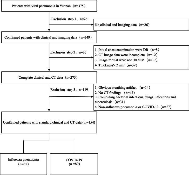
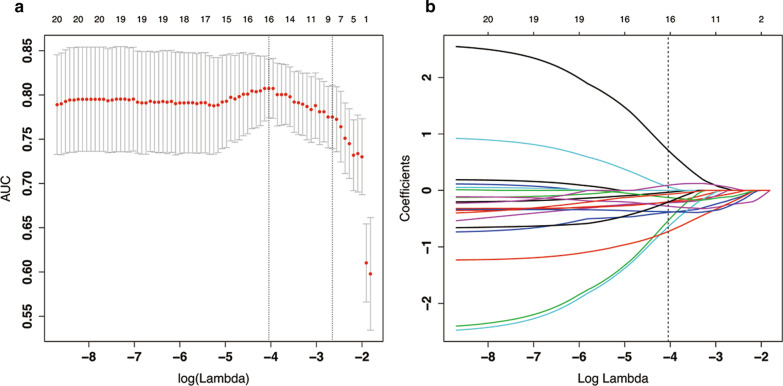
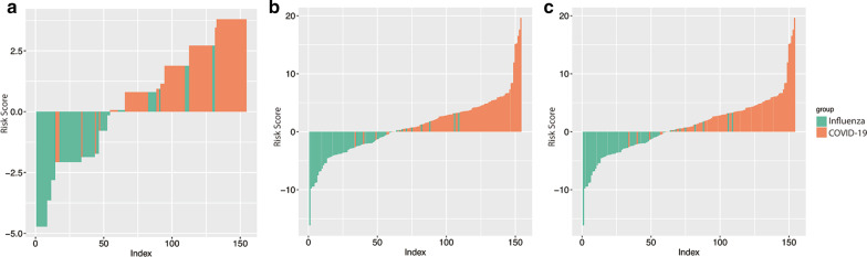
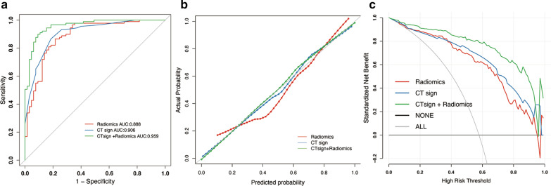
Methods: A total of 154 patients with confirmed viral pneumonia (COVID-19: 89 cases, influenza pneumonia: 65 cases) were collected retrospectively in this study. Pneumonia signs and radiomics features were extracted from the initial unenhanced chest CT images to build independent and combined models. The predictive performance of the radiomics model, CT sign model, the combined model was constructed based on the whole dataset and internally invalidated by using 1000-times bootstrap. Diagnostic performance of the models was assessed via receiver operating characteristic (ROC) analysis.

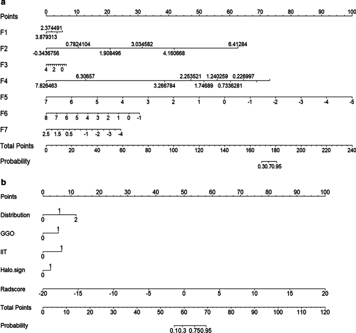
Results: The combined models consisted of 4 significant CT signs and 7 selected features and demonstrated better discrimination performance between COVID-19 and influenza pneumonia than the single radiomics model. For the radiomics model, the area under the ROC curve (AUC) was 0.888 (sensitivity, 86.5%; specificity, 78.4%; accuracy, 83.1%), and the AUC was 0.906 (sensitivity, 86.5%; specificity, 81.5%; accuracy, 84.4%) in the CT signs model. After combining CT signs and radiomics features, AUC of the combined model was 0.959 (sensitivity, 89.9%; specificity, 90.7%; accuracy, 90.3%).

Conclusions: CT-based radiomics combined with signs might be a potential method for distinguishing COVID-19 and influenza pneumonia with satisfactory performance.

Keywords: Coronavirus disease 2019; Radiomics; Viral pneumonia; X-ray computed tomography.


Conflict of interest statement

The authors had no conflicts of interest to declare in relation to this article.

Figures

Fig. 1
Fig. 1
Flow diagram of the study design and patient enrollment in the analysis
Fig. 2
Fig. 2
The radiomic feature selection using the least absolute shrinkage and selection operator (LASSO) binary logistic regression model. a Parameter (Lambda) is tuned in the LASSO model using fivefold cross-validation via maximum area under the ROC curve criteria. AUC on each fold was drawn versus log (Lambda). Vertical line was drawn at the determined optical log (Lambda) with one standard error among fivefold cross-validation, where optimal Lambda resulted in nonzero coefficients. b A coefficient profile plotted against the log (Lambda)
Fig. 3
Fig. 3
Chest CT findings of COVID-19 pneumonia. ad Male, 28 years old, 3 days after onset, axial view by CT scan. Multiple pure GGO in the upper left lobe (a, b), intralobular interstitial thickening and halo sign (c), vascular thickening and halo sign (d). E–H: Male, 25 years old, 2 days after onset, axial view by CT scan. Multiple pure GGO in the lower of both lobe (e, g), vascular thickening and halo sign (f); intralobular interstitial thickening and halo sign (h)
Fig. 4
Fig. 4
Chest CT findings of influenza pneumonia. Male, 35 years old, 4 days after onset. Multifocal GGO, partial consolidation and consolidation in both lungs (ad). Feale, 39 years old, 6 days after onset. Multifocal GGO, partial consolidation and consolidation in the right lower lungs (e, g). tree-in-bud (f), and consolidation (h)
Fig. 5
Fig. 5
The bar chart of three models for patients with COVID-19 and influenza pneumonia. a CT signs model; b radiomics model; c combined model
Fig. 6
Fig. 6
Predictive performance of the CT signs model, radiomics model and the combined model. a Receiver operating characteristic (ROC) curves. b Calibration curves. c Decision curve analysis
Fig. 7
Fig. 7
The nomogram representing radiomics model and combined model. a Radiomics model; b combined model. F1: "lbp.3D.k_ngtdm_Contrast"; F2: "lbp.3D.k_ngtdm_Strength"; F3:"lbp.3D.m2_glszm_SmallAreaEmphasis"; F4: "wavelet.LLH_ngtdm_Contrast"; F5:"wavelet.HLL_firstorder_Mean"; F6:"lbp.3D.m2_glszm_ZoneVariance"; F7: "wavelet.LHL_gldm_DependenceEntropy"; Radscore, radiomics score; GGO, ground-glass opacities; IIT, intralobular interstitial thickening. 0: negative, 1: positive in GGO and IIT, or 0: central, 1: peripheral, 2: mix in distribution

Similar articles

Cited by

References

    1. Zhu N, Zhang D, Wang W, Li X, Yang B, Song J, Zhao X, Huang B, Shi W, Lu R, et al. A novel coronavirus from patients with Pneumonia in China, 2019. N Engl J Med. 2020;382(8):727–733. doi: 10.1056/NEJMoa2001017. - DOI - PMC - PubMed
    1. Guan WJ, Ni ZY, Hu Y, Liang WH, Ou CQ, He JX, Liu L, Shan H, Lei CL, Hui DSC, et al. Clinical characteristics of coronavirus disease 2019 in China. N Engl J Med. 2020;382(18):1708–1720. - PMC - PubMed
    1. Qiu H, Wu J, Hong L, Luo Y, Song Q, Chen D. Clinical and epidemiological features of 36 children with coronavirus disease 2019 (COVID-19) in Zhejiang, China: an observational cohort study. Lancet Infect Dis. 2020;20(6):689–696. - PMC - PubMed
    1. Chen N, Zhou M, Dong X, Qu J, Gong F, Han Y, Qiu Y, Wang J, Liu Y, Wei Y, et al. Epidemiological and clinical characteristics of 99 cases of 2019 novel coronavirus pneumonia in Wuhan, China: a descriptive study. Lancet. 2020;395(10223):507–513. - PMC - PubMed
    1. Fowlkes A, Steffens A, Temte J, Lonardo SD, McHugh L, Martin K, Rubino H, Feist M, Davis C, Selzer C, et al. Incidence of medically attended influenza during pandemic and post-pandemic seasons through the Influenza Incidence Surveillance Project, 2009–13. Lancet Respir Med. 2015;3(9):709–718. - PMC - PubMed