

Reprogramming of glutamine metabolism via glutamine synthetase silencing induces cisplatin resistance in A2780 ovarian cancer cells
- PMID: 33596851
- PMCID: PMC7891143
- DOI: 10.1186/s12885-021-07879-5
Reprogramming of glutamine metabolism via glutamine synthetase silencing induces cisplatin resistance in A2780 ovarian cancer cells
Abstract
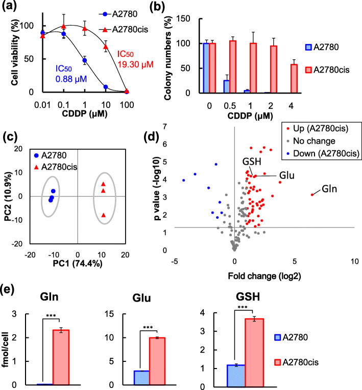
Background: Cisplatin (CDDP) significantly prolongs survival in various cancers, but many patients also develop resistance that results in treatment failure. Thus, this study aimed to elucidate the underlying mechanisms by which ovarian cancer cells acquire CDDP resistance.
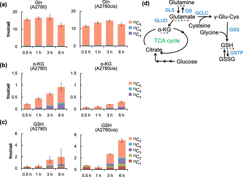
Methods: We evaluated the metabolic profiles in CDDP-sensitive ovarian cancer A2780 cells and CDDP-resistant A2780cis cells using capillary electrophoresis-time-of-flight mass spectrometry (CE-TOFMS). We further examined the expression of glutamine metabolism enzymes using real-time PCR and Western blot analyses. Cell viability was accessed using 3-(4,5-dimethylthiazol-2-yl)-2,5-diphenyltetrazolium bromide (MTT) assay.
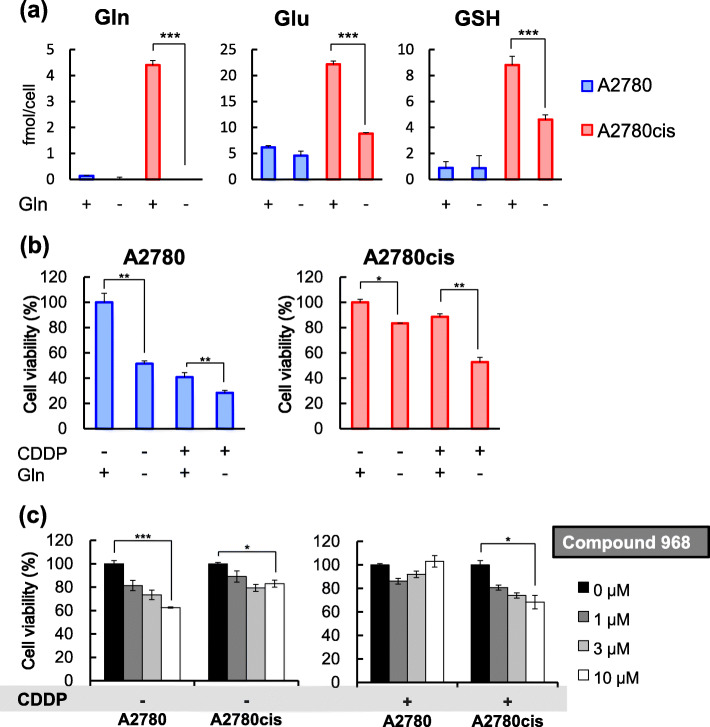
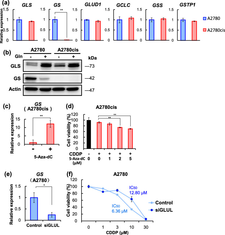
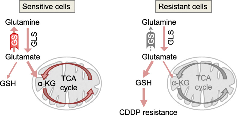
Results: The results showed that levels of glutamine, glutamate, and glutathione (GSH), a key drug resistance mediator synthesized from glutamate, were significantly elevated in A2780cis cells than those in A2780 cells. Furthermore, glutamine starvation decreased the GSH levels and CDDP resistance in A2780cis cells. Interestingly, the expression of glutamine synthetase (GS/GLUL), which synthesizes glutamine from glutamate and thereby negatively regulates GSH production, was almost completely suppressed in resistant A2780cis cells. In addition, treatment of A2780cis cells with 5-aza-2'-deoxycytidine, a DNA-demethylating agent, restored GS expression and reduced CDDP resistance. In contrast, GS knockdown in CDDP-sensitive A2780 cells induced CDDP resistance.
Conclusions: The results indicate that upregulation of GSH synthesis from glutamine via DNA methylation-mediated silencing of GS causes CDDP resistance in A2780cis cells. Therefore, glutamine metabolism could be a novel therapeutic target against CDDP resistance.
Keywords: CE-TOFMS; Cisplatin resistance; Glutamine synthetase; Metabolome; Ovarian cancer.
Conflict of interest statement
The authors declare no competing interests.
Figures
Similar articles
-
Baicalein improves the chemoresistance of ovarian cancer through regulation of CirSLC7A6.J Ovarian Res. 2023 Nov 8;16(1):212. doi: 10.1186/s13048-023-01285-0. J Ovarian Res. 2023. PMID: 37940982 Free PMC article.
-
The negative feedback between miR-143 and DNMT3A regulates cisplatin resistance in ovarian cancer.Cell Biol Int. 2021 Jan;45(1):227-237. doi: 10.1002/cbin.11486. Epub 2020 Oct 22. Cell Biol Int. 2021. PMID: 33090550
-
NCX-4040, a nitric oxide-releasing aspirin, sensitizes drug-resistant human ovarian xenograft tumors to cisplatin by depletion of cellular thiols.J Transl Med. 2008 Feb 26;6:9. doi: 10.1186/1479-5876-6-9. J Transl Med. 2008. PMID: 18302761 Free PMC article.
-
Novel factors of cisplatin resistance in epithelial ovarian tumours.Adv Med Sci. 2025 Mar;70(1):94-102. doi: 10.1016/j.advms.2025.01.005. Epub 2025 Jan 27. Adv Med Sci. 2025. PMID: 39880191 Review.
-
Cisplatin Resistance: Genetic and Epigenetic Factors Involved.Biomolecules. 2022 Sep 24;12(10):1365. doi: 10.3390/biom12101365. Biomolecules. 2022. PMID: 36291573 Free PMC article. Review.
Cited by
-
Exogenous proline enhances susceptibility of NSCLC to cisplatin via metabolic reprogramming and PLK1-mediated cell cycle arrest.Front Pharmacol. 2022 Jul 14;13:942261. doi: 10.3389/fphar.2022.942261. eCollection 2022. Front Pharmacol. 2022. PMID: 35910374 Free PMC article.
-
NMR Metabolomics of Primary Ovarian Cancer Cells in Comparison to Established Cisplatin-Resistant and -Sensitive Cell Lines.Cells. 2024 Apr 9;13(8):661. doi: 10.3390/cells13080661. Cells. 2024. PMID: 38667276 Free PMC article.
-
Ovarian Cancer and Glutamine Metabolism.Int J Mol Sci. 2023 Mar 6;24(5):5041. doi: 10.3390/ijms24055041. Int J Mol Sci. 2023. PMID: 36902470 Free PMC article. Review.
-
Unveiling the mechanisms and challenges of cancer drug resistance.Cell Commun Signal. 2024 Feb 12;22(1):109. doi: 10.1186/s12964-023-01302-1. Cell Commun Signal. 2024. PMID: 38347575 Free PMC article. Review.
-
M6 A demethylase fat mass and obesity-associated protein regulates cisplatin resistance of gastric cancer by modulating autophagy activation through ULK1.Cancer Sci. 2022 Sep;113(9):3085-3096. doi: 10.1111/cas.15469. Epub 2022 Jul 7. Cancer Sci. 2022. PMID: 35730319 Free PMC article.
References
MeSH terms
Substances
LinkOut - more resources
Full Text Sources
Other Literature Sources
Medical
Miscellaneous
