Case Reports
. 2021 Feb 17;19(1):17.
doi: 10.1186/s12969-021-00501-9.

Tibia stress injury and the imaging appearance of stress fracture in juvenile dermatomyositis: six patients' experiences

Affiliations
Case Reports

Tibia stress injury and the imaging appearance of stress fracture in juvenile dermatomyositis: six patients' experiences

Tomo Nozawa et al. Pediatr Rheumatol Online J. .

Abstract

Background: Tibial stress injuries are frequent injuries of the lower extremity and the most common causes of exercise-induced leg pain among athletes and military recruits. They sometimes occur in patients with pathological conditions of bone metabolism such as osteoporosis or rheumatoid arthritis, but there are previously no cases reported in juvenile dermatomyositis (JDM). Here we report 6 JDM patients who presented with shin pain, and the imaging appearance of tibial stress fractures or stress reactions.

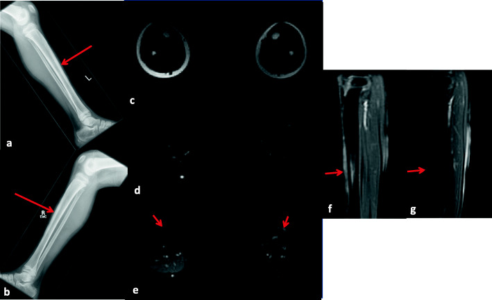
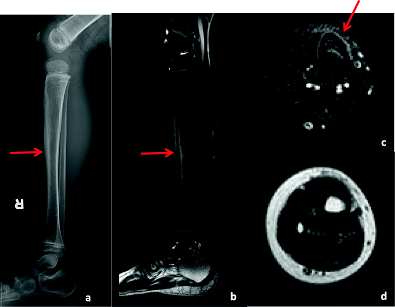
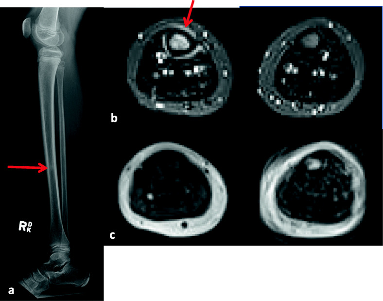
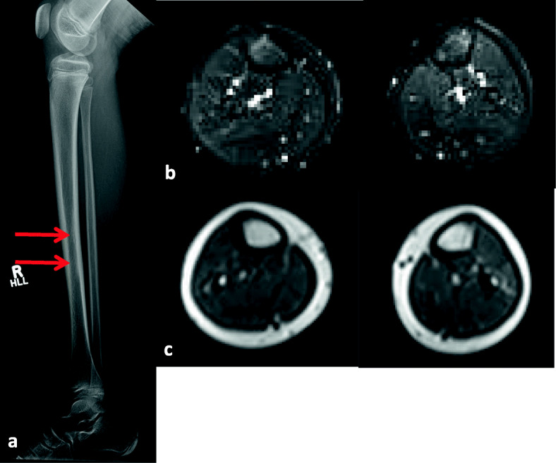
Case presentation: All 6 patients with JDM presented with shin pain or tenderness in the anterior tibia without any evidence of excessive exercise or traumatic episode. They were diagnosed with tibial stress injuries based on a combination of radiographs, three-phase bone scans, and magnetic resonance imaging (MRI), and 5 out of 6 patients had been treated with prednisone and/or methotrexate at onset of tibial stress injuries. In one patient, we could not find any abnormalities in his radiograph, but the subsequent MRI showed tibial stress reaction. In all 6 patients, the tibial stress injuries improved with only rest and/or analgesics.

Conclusion: We experienced 6 children with JDM who presented with shin pain, and who were diagnosed with tibial stress fractures or stress reactions. Their underlying disease and weakness, treatment with glucocorticoid and methotrexate, or inactivity may have resulted in these tibial injuries, and made these patients more predisposed than other children. In addition to preventing JDM patients from getting osteoporosis, we need to consider stress reactions when children with JDM complain of sudden shin pain.

Keywords: Complication; Juvenile dermatomyositis; Stress fracture; Stress reaction; Tibia; Treatment.


Conflict of interest statement

The authors declare that they have no competing interests.

Figures

Fig. 1
Fig. 1
Case 3 Lateral radiographs of the left (a) and right (b) lower extremities (a, b) show prominence of the anterior mid cortex of bilateral tibias without associated periosteal reaction or bone abnormalities (arrows) Axial T1 (c), fat saturated T2 (d) and post-gadolinium T1 (e) MR images, and sagittal post-gadolinium T1 (f, left; g, right) MR images with the lower extremities were obtained 2.5 months after the aforementioned x-rays. These images show bilateral anterior periosteal thickening, increased T2 signal intensity and enhancement noted within the bone marrow and anterior soft tissues of the mid third of both tibias (arrows), more prominent on the left than on the right
Fig. 2
Fig. 2
Case 4 Lateral radiograph of the right lower extremity (a) shows linear subperiosteal new bone formation along the mid tibial diaphysis The fat-saturated sagittal (b) and axial (c) T2 MR images of the right lower extremity obtained 4 months after the aforementioned x-rays show periosteal reaction along the mid right tibial diaphysis associated with increased signal intensity within the underlying cortex and bone marrow and at a lesser extent, anterior soft tissues (arrows). Low signal intensity is noted in the corresponding regions of the axial T1 (d) MR image. No discrete fracture line is noted
Fig. 3
Fig. 3
Case 6 (Initial observation) Lateral radiograph of the right lower extremity (a) shows a focal area of sclerosis in the mid tibial diaphysis with no associated periosteal or cortical thickening (arrow) Axial inversion recovery MR images of the thighs (b) obtained 2 weeks after the aforementioned x-rays show a focus of increased bone marrow signal in the mid right tibial diaphysis associated with a hyperintense periosteal halo on fluid sensitive images (arrow) which presents with low signal intensity on corresponding axial T1 images (c)
Fig. 4
Fig. 4
Case 6 (Follow-up) Follow-up lateral radiograph of the right lower extremity (a) obtained 3 months after the initial x-rays shows persistence of a focal area of sclerosis in the mid tibial diaphysis (arrows), increased in extent compared to previous. Subsequent axial inversion recovery (b) and T1 (c) MR images of the thighs obtained almost 1 year after the initial x-rays show interval resolution of the previously noted focus of increased bone marrow signal in the mid right tibial diaphysis

Similar articles

References

    1. Iwamoto J, Takeda T. Stress fractures in athletes: review of 196 cases. J Orthop Sci. 2003;8:273–8. doi: 10.1007/s10776-002-0632-5. - DOI - PubMed
    1. Dixon S, Newton J, Teh J. Stress fractures in the young athlete: a pictorial review. Curr Probl Diagn Radiol. 2011;40:29–44. doi: 10.1067/j.cpradiol.2009.12.001. - DOI - PubMed
    1. Devas MB. Stress fractures in children. J Bone Joint Surg Br. 1963;45:528–41. doi: 10.1302/0301-620X.45B3.528. - DOI - PubMed
    1. Keret D, Segev E, Hayek S, Lokiec F, Wientroub S. Bilateral symmetric stress fractures in a toddler. J Pediatr Orthop B. 2001;10:73–7. - PubMed
    1. Lingg GM, Soltèsz I, Kessler S, Dreher R. Insufficiency and stress fractures of the long bones occurring in patients with rheumatoid arthritis and other inflammatory diseases, with a contribution on the possibilities of computed tomography. Eur J Radiol. 1997;26:54–63. doi: 10.1016/S0720-048X(97)00039-9. - DOI - PubMed

Publication types

MeSH terms