. 2021 Feb 17;19(1):79.
doi: 10.1186/s12967-021-02744-2.

Combined lymphocyte/monocyte count, D-dimer and iron status predict COVID-19 course and outcome in a long-term care facility

Affiliations

Combined lymphocyte/monocyte count, D-dimer and iron status predict COVID-19 course and outcome in a long-term care facility

Flavia Biamonte et al. J Transl Med. .

Abstract

Background: The Sars-CoV-2 can cause severe pneumonia with multiorgan disease; thus, the identification of clinical and laboratory predictors of the progression towards severe and fatal forms of this illness is needed. Here, we retrospectively evaluated and integrated laboratory parameters of 45 elderly subjects from a long-term care facility with Sars-CoV-2 outbreak and spread, to identify potential common patterns of systemic response able to better stratify patients' clinical course and outcome.

Methods: Baseline white blood cells, granulocytes', lymphocytes', and platelets' counts, hemoglobin, total iron, ferritin, D-dimer, and interleukin-6 concentration were used to generate a principal component analysis. Statistical analysis was performed by using R statistical package version 4.0.

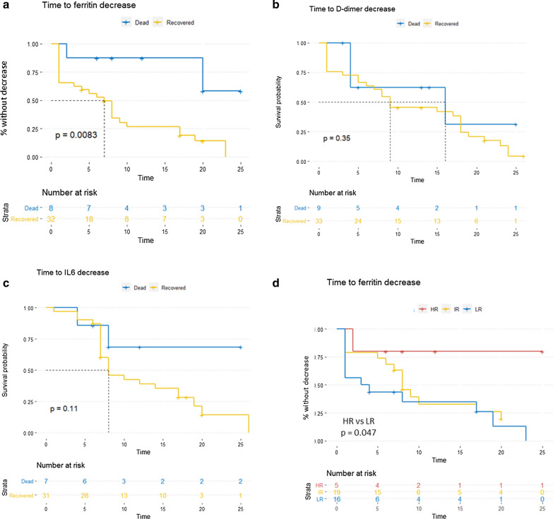
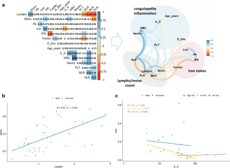
Results: We identified 3 laboratory patterns of response, renamed as low-risk, intermediate-risk, and high-risk, strongly associated with patients' survival (p < 0.01). D-dimer, iron status, lymphocyte/monocyte count represented the main markers discriminating high- and low-risk groups. Patients belonging to the high-risk group presented a significantly longer time to ferritin decrease (p: 0.047). Iron-to-ferritin-ratio (IFR) significantly segregated recovered and dead patients in the intermediate-risk group (p: 0.012).

Conclusions: Our data suggest that a combination of few laboratory parameters, i.e. iron status, D-dimer and lymphocyte/monocyte count at admission and during the hospital stay, can predict clinical progression in COVID-19.

Keywords: COVID-19; Clinical outcome; D-dimer; Iron; Long-term care facilities; Lymphocyte; Monocyte.


Conflict of interest statement

The authors declare that they have no competing interests.

Figures

Fig. 1
Fig. 1
Baseline laboratory parameters define different patterns of response to Sars-CoV-2 infection. a Clustering analysis by a principal component analysis (PCA) scatter plot. Colours and shapes respectively represent the 3 risk groups and the status of the patient (recovered or dead). b distribution of patients according to risk group and age or sex. c ANOVA and T-test results of the 6 parameters which mainly influenced patients’ clustering, according to their distribution across risk groups. Within each sub-panel, p values for ANOVA comparison (bottom) and for each pairwise comparison (top) are reported. d Patient distribution according to risk groups and dead/recovered status; on the right side, the histogram shows the comparison of iron to ferritin ratio (IFR) between recovered and dead patients within the intermediate-risk group
Fig. 2
Fig. 2
Patients with a worse outcome present a significantly longer time to ferritin decrease. ac Kaplan Meier curves reporting the time to ferritin, D-dimer and IL-6 decrease (intended as the time from hospital admission to first and stable decrease during hospitalization) respectively, stratified on dead/recovered status (p value calculated through LogRank test). d Kaplan Meier curves reporting the time to ferritin decrease stratified on risk groups (p value calculated through LogRank test comparing High-Risk vs. Low-Risk patients)
Fig. 3
Fig. 3
Correlations among lymph/monocytes count, D-dimer, and iron status affect the systemic response to SARS-CoV-2 infection. a Left: correlation plot of all variables [including derivative variables such as iron to ferritin ratio (IFR), neutrophils to lymphocytes ratio (NLR), platelets to lymphocytes ratio (PLR), monocytes to lymphocytes ratio (MLR)] to identify potential inter-variables correlations. The colour represents the direction of the correlation. Only boxes showing a significant correlation have been coloured; right, network plot showing direction and distribution of correlations between variables. Clusters of variables belonging to similar functions have been grouped. b Scatter plot representing the correlation between lymphocytes and monocytes. Remarkably, almost all the deaths are localized within the lower left quadrant of the plot. c Scatter plot showing the association between IL-6 and IFR. This correlation reaches statistical significance within the intermediate group only, further supporting the discriminating role of IFR within this subgroup
Fig. 4
Fig. 4
Association between comorbidities and baseline levels of laboratory variables. a Distribution of different laboratory values according to baseline comorbidities: left, radar-plot showing the p values of the t-test comparing each laboratory variable with each comorbidity. The distance from the center correlates with the significance of the test (the more is far the more is significant); right, box plot representing the distribution of only the significant variables in patients presenting with (1) or without (0) comorbidities. b Radar-plot reporting CVD (cardiovascular disease) associated variables: none of them presents a significantly different distribution of laboratory variables. c Distribution of COVID-19-associated deaths according to the presence or absence of malignancies at baseline

Similar articles

Cited by

References

    1. Wiersinga WJ, Rhodes A. Pathophysiology, transmission, diagnosis, and treatment of coronavirus disease 2019 (COVID-19): a review. JAMA. 2019 doi: 10.1001/jama.2020.12839. - DOI - PubMed
    1. Harrison AG, Lin T, Wang P. Mechanisms of SARS-CoV-2 transmission and pathogenesis. Trends Immunol. 2020;41(12):1100–1115. doi: 10.1016/j.it.2020.10.004. - DOI - PMC - PubMed
    1. Wang W, Xu Y, Gao R, Lu R, Han K, Wu G, Tan W. Detection of SARS-CoV-2 in different types of clinical specimens. JAMA. 2020;323(18):1843–1844. - PMC - PubMed
    1. Peng L, Liu J, Xu W, Luo Q, Chen D, Lei Z, Huang Z, Li X, Deng K, Lin B, Gao Z. SARS-CoV-2 can be detected in urine, blood, anal swabs, and oropharyngeal swabs specimens. J Med Virol. 2020;92(9):1676–1680. doi: 10.1002/jmv.25936. - DOI - PMC - PubMed
    1. Tian J, et al. Clinical characteristics and risk factors associated with COVID-19 disease severity in patients with cancer in Wuhan, China: a multicentre, retrospective, cohort study. Lancet. 2020;21(7):893–903. doi: 10.1016/S1470-2045(20)30309-0. - DOI - PMC - PubMed

MeSH terms