. 2021 Feb 17;12(1):141.
doi: 10.1186/s13287-021-02192-1.

Conditioned medium from primary cytotrophoblasts, primary placenta-derived mesenchymal stem cells, or sub-cultured placental tissue promoted HUVEC angiogenesis in vitro

Affiliations

Conditioned medium from primary cytotrophoblasts, primary placenta-derived mesenchymal stem cells, or sub-cultured placental tissue promoted HUVEC angiogenesis in vitro

Haiying Ma et al. Stem Cell Res Ther. .

Abstract

Background: As a large capillary network, the human placenta plays an important role throughout pregnancy. Placental vascular development is complex and delicate and involves many types of placental cells, such as trophoblasts, and mesenchymal stem cells. There has been no systematic, comparative study on the roles of these two groups of placental cells and the whole placental tissue in the placental angiogenesis. In this study, primary cytotrophoblasts (CTBs) from early pregnancy and primary human placenta-derived mesenchymal stem cells (hPDMSCs) from different stages of pregnancy were selected as the cell research objects, and full-term placental tissue was selected as the tissue research object to detect the effects of their conditioned medium (CM) on human umbilical vein endothelial cell (HUVEC) angiogenesis.

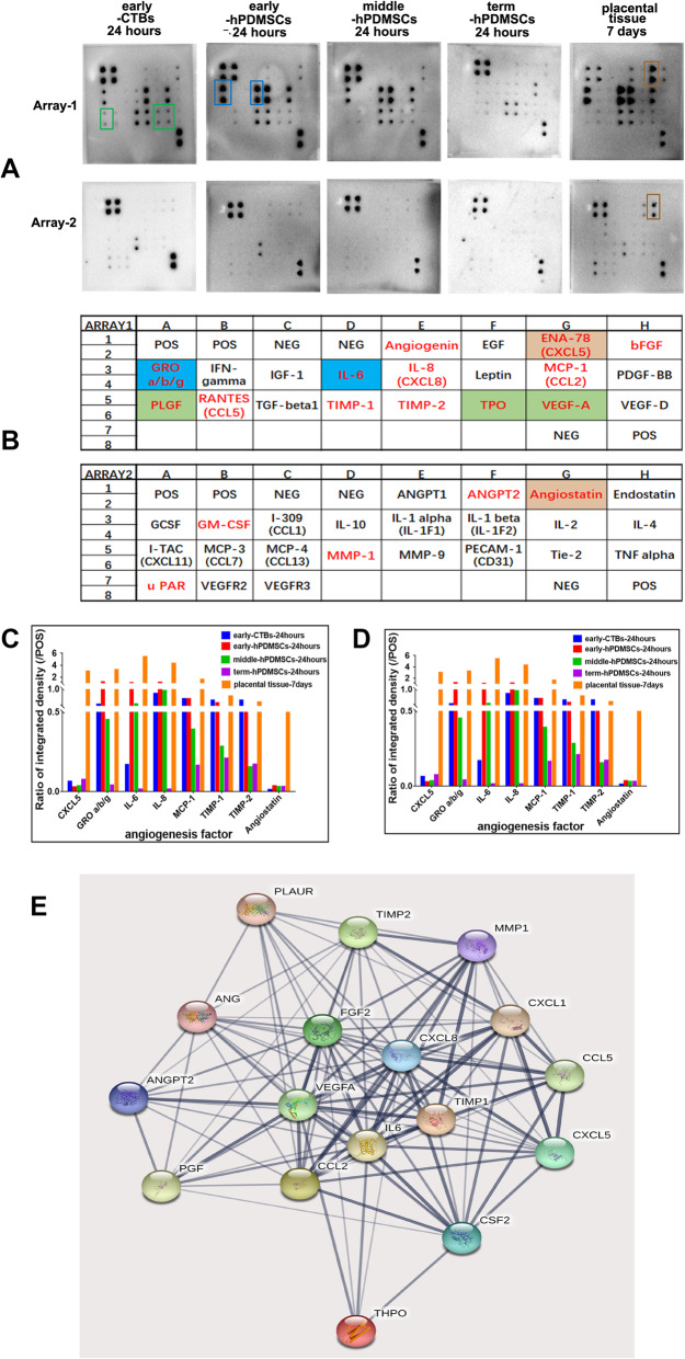
Methods: We successfully isolated primary hPDMSCs and CTBs, collected CM from these placental cells and sub-cultured placental tissue, and then evaluated the effects of the CM on a series of angiogenic processes in HUVECs in vitro. Furthermore, we measured the levels of angiogenic factors in the CM of placental cells or tissue by an angiogenesis antibody array.

Results: The results showed that not only placental cells but also sub-cultured placental tissue, to some extent, promoted HUVEC angiogenesis in vitro by promoting proliferation, adhesion, migration, invasion, and tube formation. We also found that primary placental cells in early pregnancy, whether CTBs or hPDMSCs, played more significant roles than those in full-term pregnancy. Placental cell-derived CM collected at 24 h or 48 h had the best effect, and sub-cultured placental tissue-derived CM collected at 7 days had the best effect among all the different time points. The semiquantitative angiogenesis antibody array showed that 18 of the 43 angiogenic factors had obvious spots in placental cell-derived CM or sub-cultured placental tissue-derived CM, and the levels of 5 factors (including CXCL-5, GRO, IL-6, IL-8, and MCP-1) were the highest in sub-cultured placental tissue-derived CM.

Conclusions: CM obtained from placental cells (primary CTBs or hPDMSCs) or sub-cultured placental tissue contained proangiogenic factors and promoted HUVEC angiogenesis in vitro. Therefore, our research is helpful to better understand placental angiogenesis regulation and provides theoretical support for the clinical application of placental components, especially sub-cultured placental tissue-derived CM, in vascular tissue engineering and clinical treatments.

Keywords: Angiogenesis; Conditioned medium; HUVECs; Placental tissue; Primary cytotrophoblasts; Primary human placenta-derived mesenchymal stem cells.


Conflict of interest statement

The authors declare no competing interests.

Figures

Fig. 1
Fig. 1
Characterization of primary early cytotrophoblast cells (early-CTBs) and primary human placenta-derived mesenchymal stem cells (hPDMSCs). a Morphological features of hPDMSCs isolated from human term placental tissue and passaged. The morphological features of primary hPDMSCs isolated from human full-term placental tissue (left), and the morphology features as fibroblast-like adherent cells after trypsin digestion at 0 passage (middle) and at 3rd passage (right). Scale bar 200 μm. b The cell surface phenotype of hPDMSCs was analyzed by flow cytometric analysis. The cells were positive for mesenchymal cell markers such as CD73, CD90, and CD105, and negative for hematopoietic cell markers such as CD34 and CD45. c hPDMSCs exhibited multilineage differentiation potential including endotheliocytes, osteoblasts, and adipocytes when they were cultured in the corresponding differentiation medium. The induced endothelial cells were stained with von Willebrand factor (vWF) (left), osteoblast with alkaline phosphatase (middle), and adipocytes with oil red O (right). Scale bar 100 μm. d Immunofluorescence was conducted to evaluate markers of cytotrophoblast cells in the placenta villus (left), the isolated primary early-CTBs (middle), and HTR8 cells line (right). Cytotrophoblast cells were stained by CK7 (red), mesenchymal cells by vimentin (green), and the cell nuclei were counterstained by 4′, 6-diamidino-2-phenylindole, dihydrochloride (DAPI) (blue). Scale bar 200 μm. e The early-CTBs formed multiple epithelial-like cell clones and the syncytiotrophoblast cells with the extension of the culture time. The proliferative primary CT under a light microscope (left) and the syncytiotrophoblast cells under a light microscope (middle) and immunofluorescence analyze (right). Scale bar 200 μm. g The markers of cytotrophoblasts were analyzed by flow cytometric analysis. The cells were positive for CK7 (cytotrophoblast marker) and negative for vimentin (mesenchymal cell marker)
Fig. 2
Fig. 2
The effect of conditioned medium derived from different placental cells or sub-cultured placental tissues on the proliferation of HUVECs. a The growth curve on HUVECs culturing with early-CTBs-CM obtained at different time points. b The growth curve on HUVECs culturing with early-hPDMSCs-CM obtained at different time points. c The growth curve on HUVECs culturing with middle-hPDMSCs-CM obtained at different time points. d The growth curve on HUVECs culturing with term-hPDMSCs-CM obtained at different time points. e The graph of the HUVECs number on the 6th day of proliferation assay with different placental cells CM obtained at 24 h. f The growth curve on HUVECs culturing with sub-cultured placental tissue-CM obtained at different time points. *p < 0.05 and **p < 0.01 vs control. δp < 0.05 and δδp < 0.01 vs another group within the group
Fig. 3
Fig. 3
The pro-adhesive effect of conditioned medium derived from different placental cell types or sub-cultured placental tissue on HUVECs. a Representative images of the adherent HUVECs cultured with CM derived from different placental cell types or sub-cultured placental tissue 2 h after seeding. b The graph of the adhesive effect on HUVECs by CM derived from different placental cell types obtained at different time points. c The graph of the adhesive effect on HUVECs by CM derived from different placenta cell types (early-CTBs, early-hPDMSCs; middle -hPDMSCs, and term-hPDMSCs). d The graph of the adhesive effect on HUVECs by CM obtained at different time points (6, 12, 24, 48, and 72 h). e The graph of the adhesive effect on HUVECs by CM derived from sub-cultured placental tissue obtained at different time points (1, 3, 5, 7, 10, and 14 days). *p < 0.05 and **p < 0.01 vs control. δp < 0.05 and δδp < 0.01 vs another group within the group. #p < 0.05 and ##p < 0.01 vs another group between groups, ns indicates no significant difference
Fig. 4
Fig. 4
The effect of CM derived from placental cells or sub-cultured placental tissue on the horizontal migration of HUVECs in wound healing assay. a Representative images of HUVECs both at 0 h and incubated for 8 h with CM derived from different placental cell types or sub-cultured placental tissue in wound healing assay. b The quantitative assessment of the promoting horizontal migration effect on HUVECs by CM derived from different placental cell types obtained at different time points. c The graph of the promoting horizontal migration effect on HUVECs by CM derived from different placenta cell types (early-CTBs, early-hPDMSCs; middle-hPDMSCs, and term-hPDMSCs). d The graph of the promoting horizontal migration effect on HUVECs by CM obtained at different time points (6, 12, 24, 48, and 72 h). e The graph of the promoting horizontal migration effect on HUVECs by CM derived from sub-cultured placental tissue obtained at different time points (1, 3, 5, 7, 10, and 14 days). *p < 0.05 and **p < 0.01 vs control. δp < 0.05 and δδp < 0.01 vs another group within the group. #p < 0.05 and ##p < 0.01 vs another group between groups, ns indicates no significant difference
Fig. 5
Fig. 5
The effect of CM derived from placental cells or sub-cultured placental tissue on the vertical migration of HUVECs in transwell migration assay. a Representative images of migrated HUVECs incubated for 18 h with CM derived from different placental cell types or sub-cultured placental tissue in transwell migration assay. b The quantitative assessment of the promoting vertical migration effect on HUVECs by CM derived from different placental cell types obtained at different time points. c The graph of the promoting vertical migration effect on HUVECs by CM derived from different placental cell types (early-CTBs, early-hPDMSCs; middle-hPDMSCs, and term-hPDMSCs). d The graph of the promoting vertical migration effect on HUVECs by CM obtained at different time points (6, 12, 24, 48, and 72 h). e The graph of the promoting horizontal migration effect on HUVECs by CM derived from sub-cultured placental tissue obtained at different time points (1, 3, 5, 7, 10, and 14 days). *p < 0.05 and **p < 0.01 vs control. δp < 0.05 and δδp < 0.01 vs another group within the group. #p < 0.05 and ##p < 0.01 vs another group between groups, ns indicates no significant difference
Fig. 6
Fig. 6
The effect of CM derived from placental cells or sub-cultured placental tissue on the invasion ability of HUVECs in transwell invasion assay. a Representative images of invading HUVECs incubated for 18 h with CM derived from different placental cell types or sub-cultured placental tissue in transwell invasion assay. b The quantitative assessment of the promoting invasion effect on HUVECs by CM derived from different placental cell types obtained at different time points. c The graph of the promoting invasion effect on HUVECs by CM derived from different placental cell types (early-CTBs, early-hPDMSCs; middle-hPDMSCs, and term-hPDMSCs). d The graph of the promoting invasion effect on HUVECs by CM obtained at different time points (6, 12, 24, 48, and 72 h). e The graph of the promoting invasion effect on HUVECs by CM derived from sub-cultured placental tissue obtained at different time points (1, 3, 5, 7, 10, and 14 days). *p < 0.05 and **p < 0.01 vs control. δp < 0.05 and δδp < 0.01 vs another group within the group. #p < 0.05 and ##p < 0.01 vs another group between groups, ns indicates no significant difference
Fig. 7
Fig. 7
The proangiogenic effect of CM derived from placental cells or sub-cultured placental tissue in Matrigel tube formation assay. a Representative images of the capillary-like tube structures of HUVECs incubated for 15 h with CM derived from different placental cell types or sub-cultured placental tissue in Matrigel tube formation assay. b The quantitative analysis of the total length of tube formation of HUVECs by CM derived from different placental cell types obtained at different time points. c The graph of the angiogenic effect on HUVECs by CM derived from different placental cell types (early-CTBs, early-hPDMSCs; middle-hPDMSCs, and term-hPDMSCs). d The graph of the angiogenic effect on HUVECs by CM obtained at different time points (6, 12, 24, 48, and 72 h). e The graph of the angiogenic effect on HUVECs by CM derived from sub-cultured placental tissue obtained at different time points (1, 3, 5, 7, 10, and 14 days). *p < 0.05 and **p < 0.01 vs control. δp < 0.05 and δδp < 0.01 vs another group within the group. #p < 0.05 and ##p < 0.01 vs another group between groups, ns indicates no significant difference
Fig. 8
Fig. 8
Angiogenic factor expression of placental-cell-derived CM or sub-cultured placental tissue-derived CM in human angiogenesis antibody array. a Representative images of the content of 43 conventional angiogenic factors in the CM of four types of placental cells cultured for 24 h or the CM of sub-cultured placental tissue cultured for 7 days were detected by the human angiogenesis antibody array. b The layout of 46 conventional angiogenic factors in angiogenesis antibody array membrane. c, d The histogram of the expression of angiogenic factors in the CM of four types of placental cell cultured for 24 h or the CM of placental tissue cultured for 7 days. e The analysis of protein-protein interaction of secreted proteins of the placenta. All these 17 factors could interact with others and the thickness of the connecting lines represents the strength of the interaction between factors. The red font represents all detected angiogenic factors in the placenta. The brown box represents the high content of this angiogenic factor in sub-cultured placental tissue-CM, the green box represents the high content of this angiogenic factor in early-CTBs-CM, and the blue box represents the high content of this angiogenic factor in hPDMSCs-CM

Similar articles

Cited by

References

    1. Loegl J, Nussbaumer E, Cvitic S, Huppertz B, Desoye G, Hiden U. GDM alters paracrine regulation of feto-placental angiogenesis via the trophoblast. Lab Investig. 2017;97(4):409–418. doi: 10.1038/labinvest.2016.149. - DOI - PubMed
    1. Burton GJ, Charnock-Jones DS, Jauniaux E. Regulation of vascular growth and function in the human placenta. Reproduction. 2009;138(6):895–902. doi: 10.1530/REP-09-0092. - DOI - PubMed
    1. Alfaidy N, Hoffmann P, Boufettal H, Samouh N, Aboussaouira T, Benharouga M, Feige JJ, Brouillet S. The multiple roles of EG-VEGF/PROK1 in normal and pathological placental angiogenesis. Biomed Res Int. 2014;2014:451906. doi: 10.1155/2014/451906. - DOI - PMC - PubMed
    1. Wang Y, Zhao S. Vascular biology of the placenta. San Rafael: Morgan & Claypool Life Sciences; 2010. - PubMed
    1. Borowicz PP, Arnold DR, Johnson ML, Grazul-Bilska AT, Redmer DA, Reynolds LP. Placental growth throughout the last two thirds of pregnancy in sheep: vascular development and angiogenic factor expression. Biol Reprod. 2007;76(2):259–267. doi: 10.1095/biolreprod.106.054684. - DOI - PubMed

Publication types

Substances