. 2021 Feb 17;52(1):25.
doi: 10.1186/s13567-020-00868-9.

Nanobody Nb6 fused with porcine IgG Fc as the delivering tag to inhibit porcine reproductive and respiratory syndrome virus replication in porcine alveolar macrophages

Affiliations

Nanobody Nb6 fused with porcine IgG Fc as the delivering tag to inhibit porcine reproductive and respiratory syndrome virus replication in porcine alveolar macrophages

Lu Zhang et al. Vet Res. .

Abstract

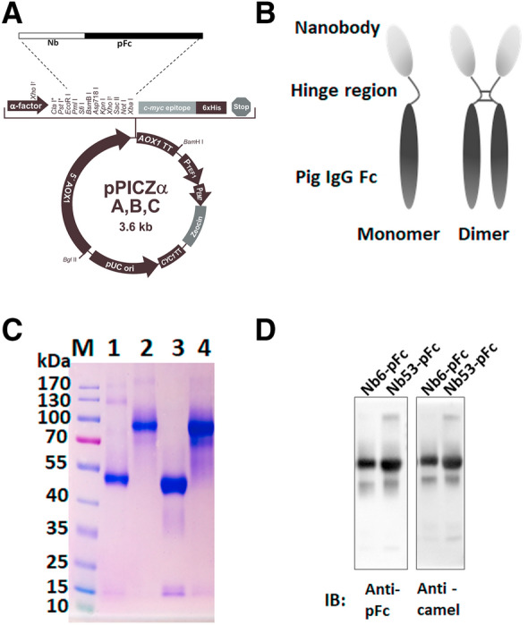
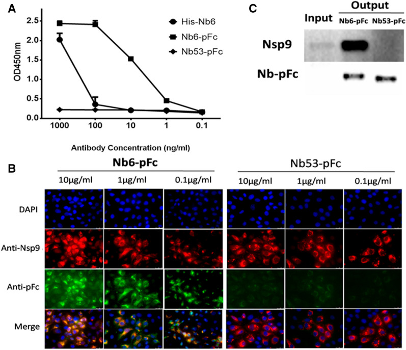
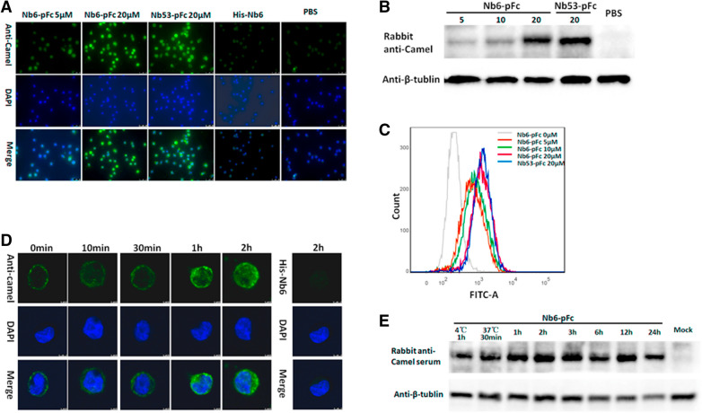
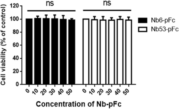
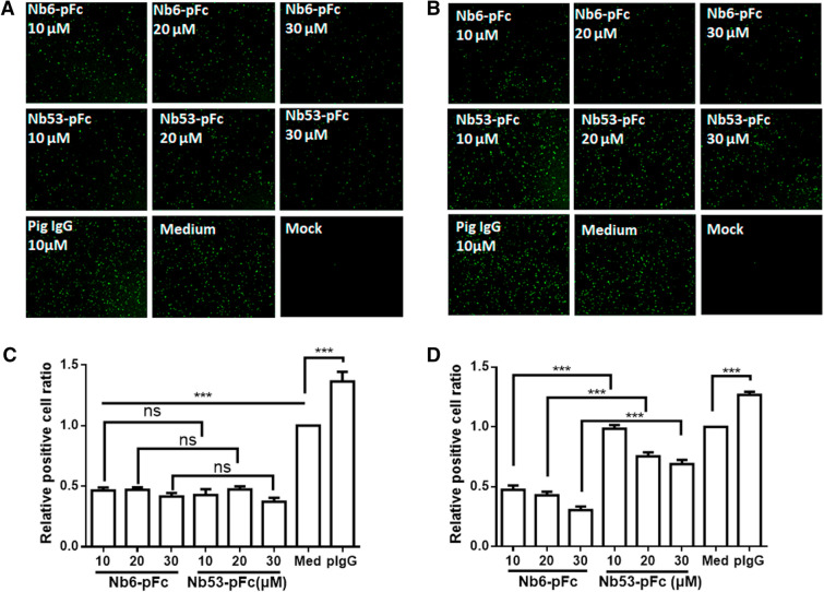
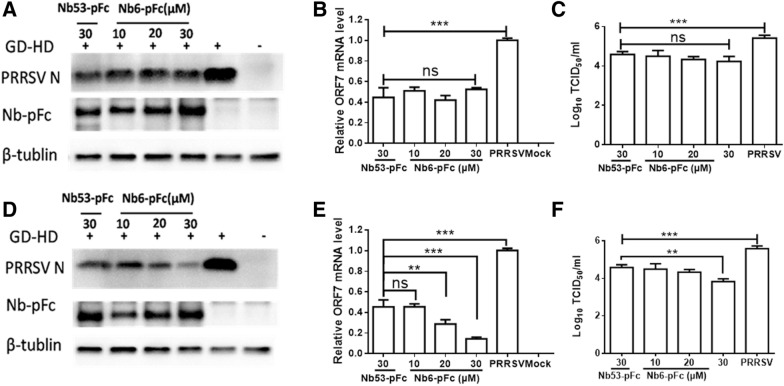
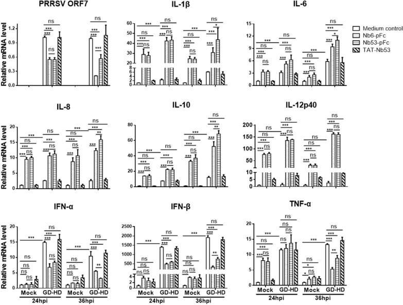
Porcine reproductive and respiratory syndrome virus (PRRSV) is a highly contagious virus that has led to enormous economic loss worldwide because of ineffective prevention and treatment. In view of their minimized size, high target specificity and affinity, nanobodies have been extensively investigated as diagnostic tools and treatments of many diseases. Previously, a PRRSV Nsp9-specific nanobody (Nb6) was identified as a PRRSV replication inhibitor. When it was fused with cell-penetrating peptide (CPP) TAT, Nb6-TAT could enter the cells for PRRSV suppression. However, delivery of molecules by CPP lack cell specificity and have a short duration of action. PRRSV has a tropism for monocyte/macrophage lineage, which expresses high levels of Fcγ receptors. Herein, we designed a nanobody containing porcine IgG Fc (Fcγ) to inhibit PRRSV replication in PRRSV permissive cells. Fcγ fused Nb6 chimeric antibody (Nb6-pFc) was assembled into a dimer with interchain disulfide bonds and expressed in a Pichia pastoris system. The results show that Nb6-pFc exhibits a well-binding ability to recombinant Nsp9 or PRRSV-encoded Nsp9 and that FcγR-mediated endocytosis of Nb6-pFc into porcine alveolar macrophages (PAM) was in a dose-dependent manner. Nb6-pFc can inhibit PRRSV infection efficiently not only by binding with Nsp9 but also by upregulating proinflammatory cytokine production in PAM. Together, this study proposes the design of a porcine IgG Fc-fused nanobody that can enter PRRSV susceptible PAM via FcγR-mediated endocytosis and inhibit PRRSV replication. This research reveals that nanobody-Fcγ chimeric antibodies might be effective for the control and prevention of monocyte/macrophage lineage susceptible pathogeneses.

Keywords: PRRSV; antiviral agents; nanobody; nanobody-pFc; permissive cell targeting.


Conflict of interest statement

The authors declare that they have no competing interests.

Figures

Figure 1
Figure 1
Construction and analysis of purified pig Fc linked nanobodies. A Design and construction of pPICZα-A-Nbs-pFc. B Speculative structural model of Nbs-pFc. C SDS-PAGE identification of purified Nbs-pFc. The predicted sizes of the pFc linked nanobodies are 80 kDa in absence of β-mercaptoethanol and 40 kDa in presence of β-mercaptoethanol. M, protein marker; lanes 1 to 4, reduced Nb6-pFc, non-reduced Nb6-pFc, reduced Nb53-pFc, non-reduced Nb53-pFc, respectively. D Western blot identification of purified pig Fc-linked nanobodies by HRP-conjugated goat anti-swine IgG and rabbit anti-camel sera.
Figure 2
Figure 2
Detection of Nb6-pFc interacted with Nsp9. A ELISA detection of binding with prokaryotic expressedNsp9. Different concentrations of Nb6-pFc interacted with Nsp9. His-Nb6 and Nb53-pFc were used as positive and negative antibody controls, respectively. Assays were performed in duplicate, and data are presented as mean ± SD. B IFA detection of binding with PRRSV Nsp9. Nb6-pFc and Nb53-pFc were detected by Alexa flour 488-conjugated goat anti-swine IgG (Green) and positive infected cells were stained by mouse anti-Nsp9 serum (Red). C MARC-145 cells were inoculated with SD16 at a MOI of 0.1 for 36 h, and Nsp9 was pulled down by Nb6-pFc and Nb53-pFc. The bound proteins were detected by Western blot using mouse anti-Nsp9 serum and HRP-conjugated goat anti-swine IgG Fc.
Figure 3
Figure 3
Cellular uptake of Nbs-pFc by PAM. A IFA, B Western blot, and C flow cytometry analyses of Nbs-pFc entry into PAM at different concentrations. PAM were treated with 0–20 μM Nbs-pFc for 2 h. D IFA and E Western blot analyses of uptake of 20 μM Nb6-pFc into PAM at different time points of incubation.
Figure 4
Figure 4
Detection of Nbs-pFc toxicity using CCK8 kits. Data are expressed as mean ± SD from five independent experiments. P values were calculated using ANOVA. P values were < 0.05 (*), < 0.01 (**), and < 0.001 (***) compared with normal cultured cells (ns: not significant).
Figure 5
Figure 5
Inhibition of rSD16/TRS2/clover replication by Nb6-pFc in PAM. Fluorescence was observed with a microscope (Leica DMI6000B Leica, Germany) at A 24 hpi and B 36 hpi, and PRRSV positive particles were calculated using Image J software and compared with the medium control group to estimate inhibition efficiency of Nb6-pFc at C 24 hpi and D 36 hpi. Data are expressed as mean ± SD from three independent tests. P values were calculated using ANOVA as < 0.05 (*), < 0.01 (**), and < 0.001 (***) compared with cells infected with PRRSV alone (ns: not significant).
Figure 6
Figure 6
HP-PRRSV GD-HD replication by Nb6-pFc in PAM. Nbs-pFc and PRRSV Nsp9 were detected at A 24 hpi and D 36 hpi by Western blot using HRP-goat anti-swine IgG and anti-PRRSV N protein mAb, respectively. Relative levels of PRRSV RNA at B 24 hpi and E 36 hpi detected by RT-qPCR using PRRSV ORF7-specific primers. The GAPDH mRNA level served as an internal reference. Progeny virus released in the cell medium was measured by TCID50 at C 24 hpi and F 36 hpi. Data are expressed as means from three independent experiments. P values were calculated using ANOVA as < 0.05 (*), < 0.01 (**), and < 0.001 (***) compared with cells infected with PRRSV alone (ns: not significant).
Figure 7
Figure 7
Regulation of cytokines by Nbs-pFc with or without PRRSV infection. PAM were innoculated with or without GD-HD (MOI of 0.01) for 1 h then treated with 30 μM of Nb6-pFc, Nb53-pFc and TAT-Nb53, and collected at 24 and 36 hpi. Relative mRNA levels of PRRSV ORF7, IL-1β, IL-6, IL-8, IL-10, IL-12p40, IFN-α, IFN-β, and TNF-α were assessed using quantitative real-time qRT-PCR. Values were normalized to the internal GAPDH control. Data are representative of three independent experiments performed in triplicate and are shown as the mean ± SD. P values were calculated using ANOVA as < 0.05 (*), < 0.01 (**), and < 0.001 (***) compared with cells infected with PRRSV alone (ns: not significant).

Similar articles

Cited by

References

    1. Li C, Tang ZR, Hu ZX, Wang YW, Yang XM, Mo FZ, Lu XL. Natural single-domain antibody-nanobody: a novel concept in the antibody field. J Biomed Nanotechnol. 2018;14:1–19. doi: 10.1166/jbn.2018.2463. - DOI - PubMed
    1. Mir MA, Mehraj U, Sheikh BA, Hamdani SS. Nanobodies: the “Magic Bullets” in therapeutics, drug delivery and diagnostics. Hum Antibodies. 2020;28:29–51. doi: 10.3233/hab-190390. - DOI - PubMed
    1. Khodabakhsh F, Behdani M, Rami A, Kazemi-Lomedasht F. Single-domain antibodies or nanobodies: a class of next-generation antibodies. Int Rev Immunol. 2018;37:316–322. doi: 10.1080/08830185.2018.1526932. - DOI - PubMed
    1. Revets H, De Baetselier P, Muyldermans S. Nanobodies as novel agents for cancer therapy. Expert Opin Biol Therapy. 2005;5:111–124. doi: 10.1517/14712598.5.1.111. - DOI - PubMed
    1. Salvador JP, Vilaplana L, Marco MP. Nanobody: outstanding features for diagnostic and therapeutic applications. Anal Bioanal Chem. 2019;411:1703–1713. doi: 10.1007/s00216-019-01633-4. - DOI - PubMed

MeSH terms