. 2021 Feb 17;13(1):45.
doi: 10.1186/s13195-021-00780-0.

CERTL reduces C16 ceramide, amyloid-β levels, and inflammation in a model of Alzheimer's disease

Affiliations

CERTL reduces C16 ceramide, amyloid-β levels, and inflammation in a model of Alzheimer's disease

Simone M Crivelli et al. Alzheimers Res Ther. .

Abstract

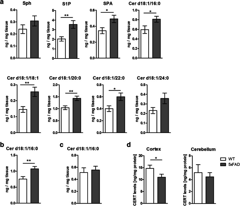
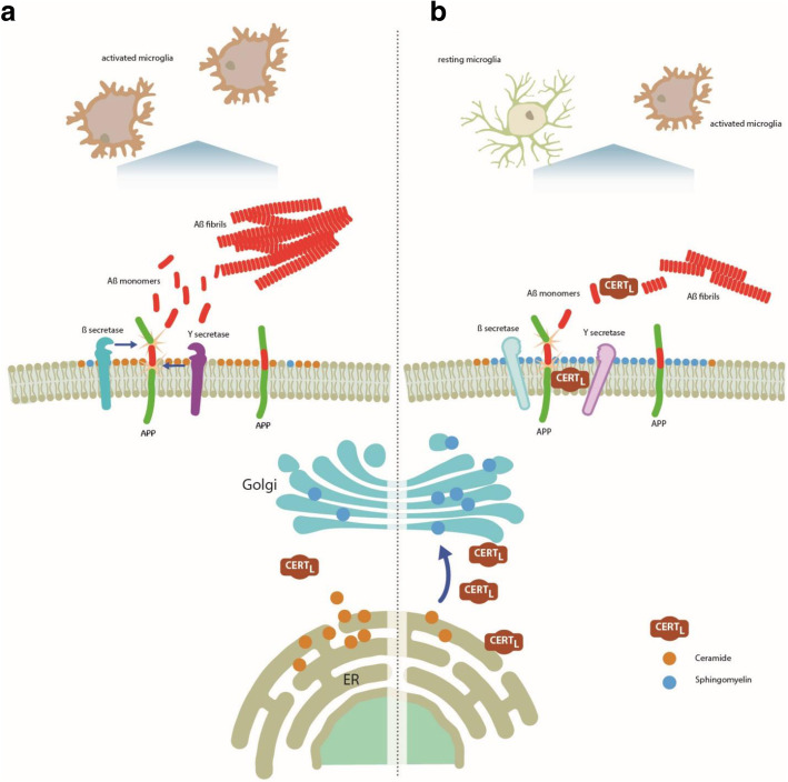
Background: Dysregulation of ceramide and sphingomyelin levels have been suggested to contribute to the pathogenesis of Alzheimer's disease (AD). Ceramide transfer proteins (CERTs) are ceramide carriers which are crucial for ceramide and sphingomyelin balance in cells. Extracellular forms of CERTs co-localize with amyloid-β (Aβ) plaques in AD brains. To date, the significance of these observations for the pathophysiology of AD remains uncertain.

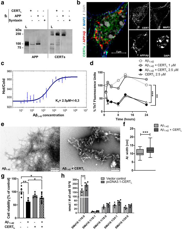
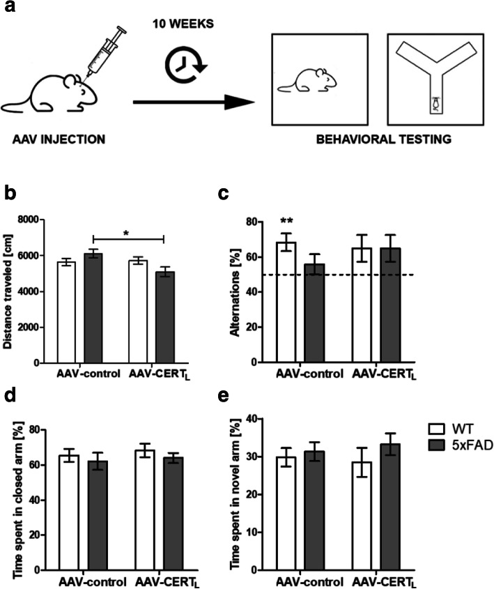
Methods: A plasmid expressing CERTL, the long isoform of CERTs, was used to study the interaction of CERTL with amyloid precursor protein (APP) by co-immunoprecipitation and immunofluorescence in HEK cells. The recombinant CERTL protein was employed to study interaction of CERTL with amyloid-β (Aβ), Aβ aggregation process in presence of CERTL, and the resulting changes in Aβ toxicity in neuroblastoma cells. CERTL was overexpressed in neurons by adeno-associated virus (AAV) in a mouse model of familial AD (5xFAD). Ten weeks after transduction, animals were challenged with behavior tests for memory, anxiety, and locomotion. At week 12, brains were investigated for sphingolipid levels by mass spectrometry, plaques, and neuroinflammation by immunohistochemistry, gene expression, and/or immunoassay.

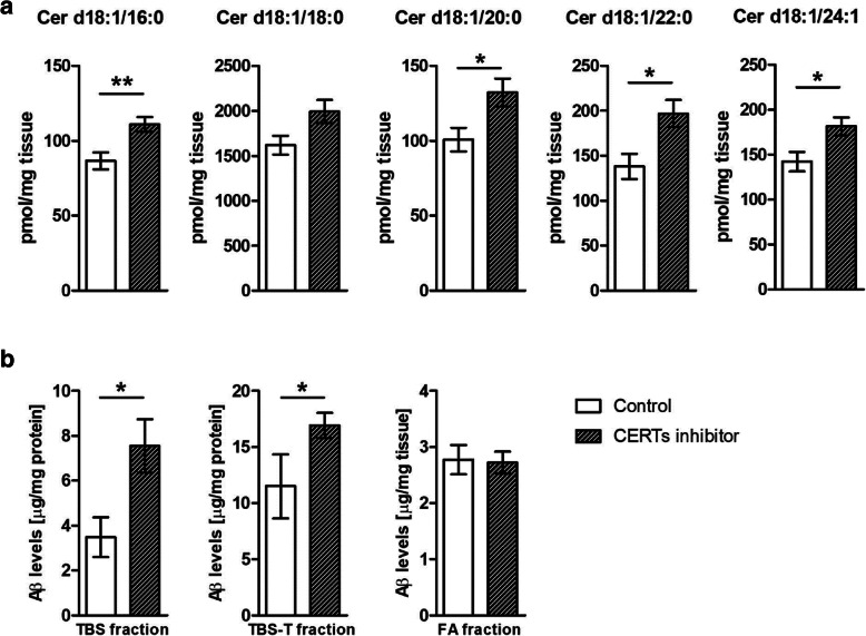
Results: Here, we report that CERTL binds to APP, modifies Aβ aggregation, and reduces Aβ neurotoxicity in vitro. Furthermore, we show that intracortical injection of AAV, mediating the expression of CERTL, decreases levels of ceramide d18:1/16:0 and increases sphingomyelin levels in the brain of male 5xFAD mice. CERTL in vivo over-expression has a mild effect on animal locomotion, decreases Aβ formation, and modulates microglia by decreasing their pro-inflammatory phenotype.

Conclusion: Our results demonstrate a crucial role of CERTL in regulating ceramide levels in the brain, in amyloid plaque formation and neuroinflammation, thereby opening research avenues for therapeutic targets of AD and other neurodegenerative diseases.

Keywords: 5xFAD; Adeno-associated virus (AAV); Alzheimer’s disease (AD); Amyloid-β plaques; Ceramide; Ceramide transporter protein (CERT); Microglia; Neuroinflammation; Sphingomyelin.


Conflict of interest statement

The authors declare that they have no competing interests.

Figures

Fig. 1
Fig. 1
CERTL binds directly to APP and Aβ and reduces Aβ aggregation and toxicity in vitro. a Protein interaction detected using co-IP of APP and CERTL in HEK-APP. The total cell lysate of HEK-APP cells (L) lane 1 and the total cell lysate of HEK-APP cells IP using APP/Aβ (lane 2), CERTL (lane 3), and syntaxin (isotype control) (lane 4) antibodies were analyzed by Western blot. APP (1) and CERTs proteins were detected. The isotype control syntaxin protein was negative. Molecular weight markers are indicated (kDa). b Immunofluorescent staining showing co-localization of CERTs and APP/Aβ in primary neurons isolated from 5xFAD brains. Neuronal marker MAP 2 was used to immuno-label neuronal cells. DAPI was used for nuclei staining. Scale bar 5 μm. c CERTL and Aβ1–42 interaction was measured by microscale thermophoresis. The dissociation constant (Kd) calculated was 2.5 ± 0.3 μM. d Percentage of Thioflavin T (ThT) fluorescence intensity to detect Aβ1–42 20 μM aggregation in the absence or presence of recombinant CERTL 1 or 2.5 μM at different time points. Each data point represents the percentage of mean fluorescent intensity of three wells (repeated measures ANOVA; Dunnett’s multiple comparisons test **p < 0.01, ***p < 0.001). e, f TEM analysis of 2 μM Aβ1–42 aggregation in the absence and presence of 0.1 μM CERTL showed a different aggregation pattern quantified by Aβ width (Student’s t test ***p < 0.001). g Measurement of cell metabolic activity of SH-SY5Y by MTT assay in cells incubated with medium alone (control) or medium containing 10 μM Aβ1–42, Aβ1–42, and 1 μM CERTL, or CERTL alone for 24 h. Graph bar expressed as means ± S.E.M % of control N = 5–10 (one-way ANOVA, Bonferroni correction *p < 0.05; **p < 0.01). h SM d18:1/16:0, SM d18:1/18:1, SM d18:1/18:0, SM d18:1/24:1, and SM d18:1/24:0 measured by HPLC-MS/MS in N2a cells after 48-h transfection with vector control or pcDNA-CERTL. Graph bar expressed as means ± S.E.M % of control N = 4/group (one-way ANOVA, Holm-Sidak’s multiple comparisons test ***p < 0.001)
Fig. 2
Fig. 2
Ceramide levels are increased in 5xFAD compared to WT animals depending on brain region and acyl chain length while CERT levels are reduced. Sphingolipids levels were measured in the hippocampus (a), cortex (b), and cerebellum (c) by HPLC-MS/MS of WT and 5xFAD mice. Sphingolipids were classified based on acyl chain number of carbons (Sph, S1P, SPA, Cer d18:1/16:0, Cer d18:1/18:1, Cer d18:1/20:0, Cer d18:1/22:0, and Cer d18:1/24:0). CERT was quantified by ELISA in protein extract of cortex and cerebellum of WT and 5xFAD animals. Bars represent the mean ± S.E.M per group N = 11–12 (Student’s t test *p < 0.05; **p < 0.01)
Fig. 3
Fig. 3
AAV-mediated neuronal expression of CERTL in mouse brain. a The recombinant genomes of the two AAV-2 vectors. ➔ Abbreviations: From left to right, ITR, inverted terminal repeats; hSYN1, human synapsin 1 gene promoter; CERTL, cDNA sequence coding for ceramide transfer protein long isoform (hCERTL, 1875 bp NP_005704.1); IRES, internal ribosome sequence for translation initiation; EGFP, cDNA coding for enhanced green fluorescent protein (GFP); WPRE, woodchuck hepatitis virus posttranscriptional control element; bGH, bovine growth hormone gene-derived polyadenylation site; TB, synthetic transcription blocker. b Representative images of immunofluorescent staining of cortical brain area from 5xFAD animals treated with AAV-control or AAV-CERTL. Section was co-stain for CERTs protein (green), neuronal marker NeuN (red). Scale bar 200 μM and 50 μM. c Western blot showing band intensities of CERTs and GAPDH. d Relative quantification of CERTs levels normalized to GAPDH in cortical protein extract from WT and 5xFAD animals treated with AAV-control or AAV-CERTL. Bars represent mean one representative experiment with 5–7 samples per group (Mann–Whitney test, *p < 0.05)
Fig. 4
Fig. 4
No behavioral abnormalities 10 weeks after injection of AAV-CERTL. a The effects of CERTL over-expression were investigated in 30 5xFAD and 30 WT males. Mice were bilaterally injected at 12–13 weeks of age with AAV-CERTL or AAV-control particles at the dose 1.12 × 10E8 transducing unit (t.u.). Starting at week 22 of age, animals were challenged with the following behavioral tests: open field (OF) for locomotion activity, alternate Y-maze (AYM) and spatial Y-maze (SYM) for spatial memory, and elevated zero-maze (EZM) for anxiety. b Locomotion expressed as distance traveled in OF task. c The graph shows the results of the working memory in the AYM task as a percentage of correct alternation in the first four triads. Percentage were compared to 50% chance levels (one sample t test **p < 0.01). d Anxiety was assessed, measuring the percentage of time spent in the closed arm in EZM. e Memory was measured in SYM expressed as a percentage of time spent in the novel arm. Bars represent the means ± S.E.M per group N = 10–20 (two-way ANOVA, interaction effect F = 4.170 p = 0.0463, LSD, *p < 0.05)
Fig. 5
Fig. 5
AAV-CERTL reduces Cer d18:1/16:0 and increases sphingomyelin species in the cortex. Sphingolipids levels were measured in the cortex by HPLC-MS/MS. Ceramides were classified based on acyl chain number of carbons (Cer d18:1/16:0, Cer d18:1/18:0, Cer d18:1/18:1, Cer d18:1/20:0, Cer d18:1/22:0, and Cer d18:1/24:1) as well as sphingomyelin (SM d18:1/16:0, SM d18:1/18:0, SM d18:1/18:1, SM d18:1/20:0, SM d18:1/22:0, and SM d18:1/24:1). Ceramide levels were expressed as ng/mg tissue, while sphingomyelins were expressed as pmol/mg tissue. Bars represent the mean ± S.E.M per group N = 5–12 (two-way ANOVA, LSD, significant effects, *p < 0.05; **p < 0.01; ***p < 0.001)
Fig. 6
Fig. 6
Neuronal increase of CERTL reduces Aβ by decreasing APP cleavage. a Representative photomicrographs of sagittal brain sections imaging the motor sensory cortex (M1 and M2) stained for nuclei in blue and Aβ plaques in green. All photomicrographs were exposed and processed identically. Scale bar represents 200 and 50 μm (from right to left). b Immunofluorescent quantification of plaques measured by the percentage of area, plaques counts/mm2. c Frequency distribution of plaques based on size (10–25 μm) (error bars represent ± SEM of 4–6 animals per experimental condition, ANOVA, Bonferroni correction, significant effects, *p < 0.05; **p < 0.01). d Aβ quantification in three extraction buffers, BS, TBS-T, and formic acid (FA) by ELISA showed that Aβ was significantly reduced in the soluble fractions in the cortex but not in the insoluble fraction (Student’s t test *p < 0.05). e Western blot analysis of TBS cortex homogenate stained with 6E10 antibody showed that ratios of amyloid Aβ/FL-APP and Aβ/CTFβ are reduced while CTFβ/FL-APP is increased in AAV-CERTL-treated animals while CTFβ/FL-APP is increased. Error bars represent ± SEM of 5 animals per experimental condition (Student’s t test *p < 0.05; **p < 0.01) (full length amyloid precursor protein = FL-APP; amyloid-β peptide = Aβ; C-terminal fragment β = CFTβ). Western blot membranes are shown in Supplementary Figure 5
Fig. 7
Fig. 7
CERTs inhibitor increases Cer and Aβ levels in the brain of transgenic AD mice. a Sphingolipid levels were measured in the cortex by HPLC-MS/MS. Ceramides were classified based on acyl chain number of carbons (Cer d18:1/16:0, Cer d18:1/18:0, Cer d18:1/22:0, and Cer d18:1/24:1) and levels were expressed as pmol/mg tissue. Bars represent the mean ± S.E.M per group N = 10–12 (Student’s t test *p < 0.05) Aβ quantification in hippocampus homogenate extracted in three buffers, TBS, TBS-T, and formic acid (FA) by enzyme-linked immunoassays. Means of each fraction were compared with unpaired t test (control N = 10; HPA-12 N = 13; *p < 0.05)
Fig. 8
Fig. 8
AAV-CERTL reduces microglia reactivity to Iba1 and CD86 expression levels but has no significant effect on GFAP immunoreactivity in the cortex of 5xFAD mice. a Representative photomicrograph of Iba1 staining in the cortical motor sensory region of 5xFAD animals treated with AAV-control or AAV-CERTL (scale bar 50 μm). b Densitometric analysis of Iba1 staining represented as a percentage of the area (AAV-control n = 6 and AAV-CERTL n = 4 for WT and 5xFAD groups). c Densitometric analysis of Iba1 staining represented as number of positive Iba1 cells/mm2 (AAV-control n = 6 and AAV-CERTL n = 4 for 5xFAD groups). d Length of microglia ramification and sphericity per cell in AAV-control or AAV-CERTL. Morphological analysis was performed on 3–5 pictures/group. e Illustrations of the microglia morphological analysis applied to a fluorescent photomicrograph captured with × 60 objective with a single cell cropped to show details. Scale bar = 20. f Analysis of gene expression of membrane markers CD86 (4–5 number of animals per group). g Representative photomicrographs of GFAP staining in the cortical motor sensory region of 5xFAD animals treated with AAV-control or AAV-CERTL (scale bar 50 μm). h Densitometric analysis of GFAP staining represented as a percentage of the area (AAV-control n = 6 and AAV-CERTL n = 4 for WT and 5xFAD groups). Bars represent the mean ± S.E.M per group (two-way ANOVA, LSD, significant effects, *p < 0.05; **p < 0.01)
Fig. 9
Fig. 9
Schematic model of CERTL action in AD. a CERTL concentration is decreased in AD neuronal cells. Consequently, the transport of Cer to the Golgi is impaired and Cer accumulates in the cell. Cer elevation stabilizes and favors the secretases activity. The amyloidogenic APP processing is favored and Aβ is produced. The neighboring microglia changes the resting status to activate. b By overexpressing CERTL, the physiological transfer of Cer from the ER to the Golgi is restored favoring SM synthesis, which is intensified. The reduction of Cer levels in neuronal cells diminished secretases activity, reducing Aβ biogenesis. The interaction between CERTL and APP may be important in stabilizing APP in the membrane and in protecting APP from secretase activity. Furthermore, CERTL affects Aβ fibrilization by organizing Aβ into less neurotoxic aggregates that may be cleared from the brain more easily and reduces the number of activated microglia

References

    1. Rogers J, Morrison JH. Quantitative morphology and regional and laminar distributions of senile plaques in Alzheimer’s disease. J Neurosci. 1985;5(10):2801–2808. doi: 10.1523/JNEUROSCI.05-10-02801.1985. - DOI - PMC - PubMed
    1. Kawasaki H, Murayama S, Tomonaga M, Izumiyama N, Shimada H. Neurofibrillary tangles in human upper cervical ganglia. Morphological study with immunohistochemistry and electron microscopy. Acta Neuropathol. 1987;75(2):156–159. doi: 10.1007/BF00687076. - DOI - PubMed
    1. Wisniewski HM, Kozlowski PB. Evidence for blood-brain barrier changes in senile dementia of the Alzheimer type (SDAT) Ann N Y Acad Sci. 1982;396:119–129. doi: 10.1111/j.1749-6632.1982.tb26848.x. - DOI - PubMed
    1. Krabbe G, Halle A, Matyash V, Rinnenthal JL, Eom GD, Bernhardt U, et al. Functional impairment of microglia coincides with Beta-amyloid deposition in mice with Alzheimer-like pathology. PLoS One. 2013;8(4):e60921. doi: 10.1371/journal.pone.0060921. - DOI - PMC - PubMed
    1. Crivelli SM, Giovagnoni C, Visseren L, Scheithauer AL, de Wit N, den Hoedt S, et al. Sphingolipids in Alzheimer’s disease, how can we target them? Adv Drug Deliv Rev. 2020. - PubMed

Publication types