. 2021 Feb 17;3(1):19.
doi: 10.1186/s42523-021-00078-4.

The dysbiosis of ovine foot microbiome during the development and treatment of contagious ovine digital dermatitis

Affiliations

The dysbiosis of ovine foot microbiome during the development and treatment of contagious ovine digital dermatitis

J S Duncan et al. Anim Microbiome. .

Abstract

Background: Contagious Ovine Digital Dermatitis (CODD) is an emerging and common infectious foot disease of sheep which causes severe welfare and economic problems for the sheep industry. The aetiology of the disease is not fully understood and control of the disease is problematic. The aim of this study was to investigate the polybacterial aetiopathogenesis of CODD and the effects of antibiotic treatment, in a longitudinal study of an experimentally induced disease outbreak using a 16S rRNA gene amplicon sequencing approach.

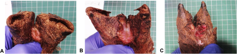
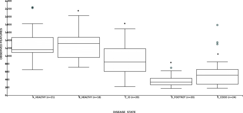
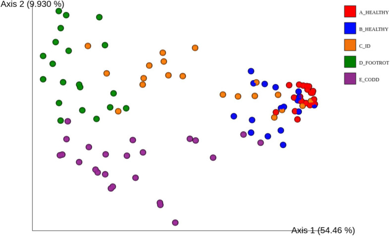
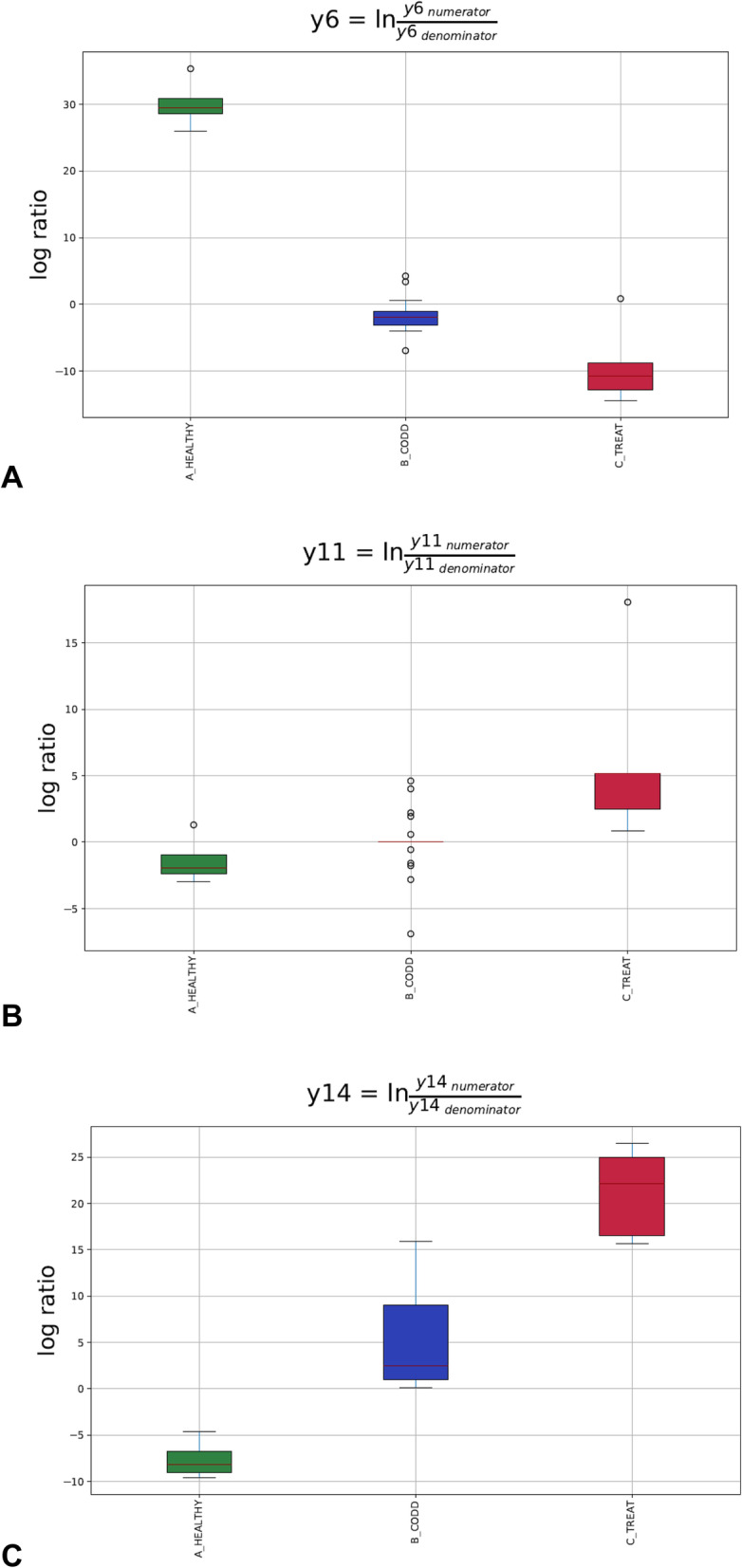
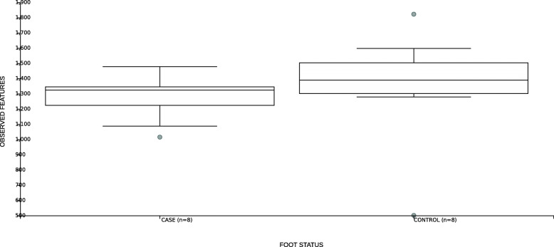
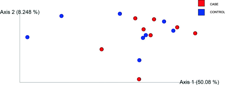
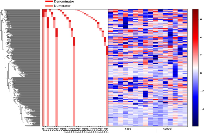
Results: CODD was induced in 15/30 experimental sheep. During the development of CODD three distinct phenotypic lesion stages were observed. These were an initial interdigital dermatitis (ID) lesion, followed by a footrot (FR) lesion, then finally a CODD lesion. Distinct microbiota were observed for each lesion in terms of microbial diversity, clustering and composition. Porphyromonadaceae, Family XI, Veillonellaceae and Fusobacteriaceae were significantly associated with the diseased feet. Veillonellaceae and Fusobacteriaceae were most associated with the earlier stages of ID and footrot rather than CODD. Following antibiotic treatment of the sheep, the foot microbiota showed a strong tendency to return to the composition of the healthy state. The microbiota composition of CODD lesions collected by swab and biopsy methods were different. In particular, the Spirochaetaceae family were more abundant in samples collected by the biopsy method, suggesting that these bacteria are present in deeper tissues of the diseased foot.

Conclusion: In this study, CODD presented as part of a spectrum of poly-bacterial foot disease strongly associated with bacterial families Porphyromonadaceae, Family XI (a family in Clostridiales also known as Clostridium cluster XI), Veillonellaceae and Fusobacteriaceae which are predominately Gram-negative anaerobes. Following antibiotic treatment, the microbiome showed a strong tendency to return to the composition of the healthy state. The composition of the healthy foot microbiome does not influence susceptibility to CODD. Based on the data presented here and that CODD appears to be the severest end stage of sheep infectious foot disease lesions, better control of the initial ID and FR lesions would enable better control of CODD and enable better animal welfare.

Keywords: CODD; Footrot; Lameness; Microbiome; Sheep.


Conflict of interest statement

The authors declare that they have no competing interests.

Figures

Fig. 1
Fig. 1
Sheep foot lesions observed during the development of CODD. a Interdigital dermatitis lesion, b footrot lesion, c CODD lesion
Fig. 2
Fig. 2
Box and whisker plots of alpha diversity as measured by observed ASVs for the different stages of CODD lesion development. * represents p < 0.05
Fig. 3
Fig. 3
Principle Coordinate Analysis (PCoA) plot showing differences in weighted UniFrac distances at different stages of CODD lesion development. ANOSIM for disease state overall (ANOSIM R statistic =0.64329, p = 0.001). Pairwise ANOSIM tests between microbiota at each phenotypic lesion stage (A_Healthy, B_Healthy, C_ID, D_Footrot, E_CODD) (p = 0.001)
Fig. 4
Fig. 4
A dendogram heatmap showing log10 abundance of ASVs in the foot microbiota at different stages of CODD lesion development
Fig. 5
Fig. 5
Gneiss analysis log10 ratio balances significantly different (p < 0.05) between stages of CODD lesion development. a balance y0, b balance y1, c balance y2, d balance y3, e balance y4(a), f balance y5, g balance y6, h balance y14. A lower log10 ratio shows a shift in the balance toward denominator ASVs whilst a higher log10 ratio shows a shift towards numerator ASVs as visually represented in Fig. 4
Fig. 6
Fig. 6
A taxa plot showing relative abundance of the 10 most abundant bacterial families (identified from the Healthy and Diseased ASV groups) in the microbiomes of the different diseased states (A_Healthy, B_Healthy, C_ID, D_Footrot and E_CODD)
Fig. 7
Fig. 7
Box and whisker plots of alpha diversity as measured by observed ASVs for samples taken from healthy (A_Healthy), CODD affected (B_CODD), antibiotic treated (C_Treated) sheep’s feet
Fig. 8
Fig. 8
Principle Coordinate Analysis (PCoA) plot showing differences in weighted UniFrac differences of samples from healthy (A_Healthy), CODD affected (B_CODD), antibiotic treated (C_Treated) sheep’s feet. ANOSIM tests show beta diversity was significantly different overall three states (R test statistic 0.825846, p = 0.001), Pairwise ANOSIM tests between microbiota at each lesion type (A_Healthy, B_CODD, C_Treated (p < 0.05)
Fig. 9
Fig. 9
Dendogram heatmap showing log10 abundance of ASV in the foot microbiota of samples from healthy (A_Healthy), CODD affected (B_CODD) and antibiotic treated (C_Treated) sheep’s feet
Fig. 10
Fig. 10
Gneiss analysis Log10 ratio balances whose log10 ratio balance was significantly different (p < 0.05) between microbiomes of samples taken from healthy (A_Healthy), CODD affected (B_CODD) and antibiotic treated (C_Treated) sheep’s feet. A lower log10 ratio shows a shift in the balance toward denominator ASVs whilst a higher log10 ratio shows a shift towards numerator ASVs as visually represented in Fig. 9
Fig. 11
Fig. 11
Box and whisker plot of alpha diversity as measured by observed ASVs for samples taken from healthy sheep’s feet that did (case) or did not (control) develop CODD
Fig. 12
Fig. 12
Principle Coordinate Analysis (PCoA) plot showing differences in weighted UniFrac distances of samples taken from healthy sheep’s feet that did (case) or did not (control) develop CODD in the study. Pairwise ANOSIM tests no difference between case and control samples (R test statistic = − 803, p = 0.803)
Fig. 13
Fig. 13
Dendogram heatmap showing log10 abundance of ASV in the foot microbiota of samples taken from healthy sheep’s feet that did (case) or did not (control) develop CODD in the study
Fig. 14
Fig. 14
A taxa bar plot showing relative abundance of bacterial families in the microbiome of the ovine foot affected by CODD in samples obtained by lesion biopsy compared with lesion swab

References

    1. Harwood DG, Cattell JH, Lewis CJ, Naylor R. Virulent footrot in sheep. Vet Rec Lett. 1997;140(26):687. - PubMed
    1. Dickins A, Clark CCA, Kaler J, Ferguson E, O’Kane H, Green LE. Factors associated with the presence and prevalence of contagious ovine digital dermatitis: a 2013 study of 1136 random English sheep flocks. Prev Vet Med. 2016;130:86–93. - PubMed
    1. Angell JW, Duncan JS, Carter SD, Grove-White DH. Farmer reported prevalence and factors associated with contagious ovine digital dermatitis in Wales: a questionnaire of 511 sheep farmers. Prev Vet Med. 2014;113(1):132–138. - PubMed
    1. Sayers G, Marques PX, Evans NJ, O'Grady L, Doherty ML, Carter SD, et al. Identification of spirochetes associated with contagious ovine digital dermatitis. J Clin Microbiol. 2009;47(4):1199–1201. - PMC - PubMed
    1. Tegtmeyer PC, Staton GJ, Evans NJ, Rohde J, Punsmann TM, Ganter M. First cases of contagious ovine digital dermatitis in Germany. Acta veterinaria Scandinavica. 2020;62(1):46. - PMC - PubMed

LinkOut - more resources