. 2021 Apr 12;95(9):e02208-20.
doi: 10.1128/JVI.02208-20. Print 2021 Apr 12.

Interferon Regulatory Factor 3 Supports the Establishment of Chronic Gammaherpesvirus Infection in a Route- and Dose-Dependent Manner

Affiliations

Interferon Regulatory Factor 3 Supports the Establishment of Chronic Gammaherpesvirus Infection in a Route- and Dose-Dependent Manner

K E Johnson et al. J Virol. .

Abstract

Gammaherpesviruses are ubiquitous pathogens that establish lifelong infections and are associated with several malignancies, including B cell lymphomas. Uniquely, these viruses manipulate B cell differentiation to establish long-term latency in memory B cells. This study focuses on the interaction between gammaherpesviruses and interferon regulatory factor 3 (IRF-3), a ubiquitously expressed transcription factor with multiple direct target genes, including beta interferon (IFN-β), a type I IFN. IRF-3 attenuates acute replication of a plethora of viruses, including gammaherpesvirus. Furthermore, IRF-3-driven IFN-β expression is antagonized by the conserved gammaherpesvirus protein kinase during lytic virus replication in vitro In this study, we have uncovered an unexpected proviral role of IRF-3 during chronic gammaherpesvirus infection. In contrast to the antiviral activity of IRF-3 during acute infection, IRF-3 facilitated establishment of latent gammaherpesvirus infection in B cells, particularly, germinal center and activated B cells, the cell types critical for both natural infection and viral lymphomagenesis. This proviral role of IRF-3 was further modified by the route of infection and viral dose. Furthermore, using a combination of viral and host genetics, we show that IRF-3 deficiency does not rescue attenuated chronic infection of a protein kinase null gammaherpesvirus mutant, highlighting the multifunctional nature of the conserved gammaherpesvirus protein kinases in vivo In summary, this study unveils an unexpected proviral nature of the classical innate immune factor, IRF-3, during chronic virus infection.IMPORTANCE Interferon regulatory factor 3 (IRF-3) is a critical component of the innate immune response, in part due to its transactivation of beta interferon (IFN-β) expression. Similar to that observed in all acute virus infections examined to date, IRF-3 suppresses lytic viral replication during acute gammaherpesvirus infection. Because gammaherpesviruses establish lifelong infection, this study aimed to define the antiviral activity of IRF-3 during chronic infection. Surprisingly, we found that, in contrast to acute infection, IRF-3 supported the establishment of gammaherpesvirus latency in splenic B cells, revealing an unexpected proviral nature of this classical innate immune host factor.

Keywords: B cell responses; IRF-3; chronic infection; gammaherpesvirus; interferon.


Figures

FIG 1
FIG 1
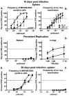
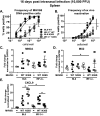
IRF-3 promotes the establishment of latent gammaherpesvirus infection. C57BL/6J (BL6) and IRF-3−/− mice were intranasally infected with 1,000 PFU of MHV68. At 16 or 28 days postinfection, limiting dilution assays were used to define frequencies of MHV68 DNA-positive cells (A and E) and MHV68 reactivation (B and F) in splenocytes pooled from 3 to 5 mice/experimental group. In the limiting dilution assays presented in the manuscript, the dotted line is drawn at 62.5% and the x coordinate of intersection of this line with the sigmoid graph represents and inverse of the frequency of positive events. Levels of preformed lytic virus in splenocytes (C) and lungs (D) at 16 days postinfection. Data were pooled from 2 to 4 independent experiments.
FIG 2
FIG 2
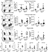
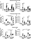
IRF-3 expression does not affect gammaherpesvirus-driven B cell differentiation. Mice of indicated genotypes were mock infected or infected as for Fig. 1. At 16 days postinfection, splenocytes were analyzed via flow cytometry. Germinal center B cells were identified as GL7+ CD95+ cells pregated on B220+ cells (representative shown in A) and expressed as frequency (B) and total cell number (C). T follicular helper cells were identified as PD1+ CXCR5+ cells pregated on CD3+ CD4+ cells (representative shown in D) and expressed as frequency (E) and total cell number (F). Plasma cells were identified as IgD-IRF4+ cells pregated on B220int/+ GL7 cells (representative shown in G) and further identified as GL7, expressed as frequency (H) and total cell number (I). (J to M) Sera collected from mice at 16 days postinfection or after mock treatment were subjected to ELISA to determine levels of indicated circulating antibodies. Each symbol represents result for an individual mouse, and data from 2 to 4 independent experiments were pooled. ***, P < 0.001.
FIG 3
FIG 3
IRF-3 facilitates MHV68 infection of germinal center/activated B cells. BL6 and IRF-3−/− mice were intranasally infected with 1,000 PFU of MHV68.ORF73βla. At 16 days postinfection, splenocytes were pooled from 3 to 5 mice/genotype, and β-lactamase activity in B220+ (A) and B220+ GL7+ (B) splenocytes was assessed by flow cytometry using fluorescence of the cleaved CCF2 β-lactamase substrate. Images representative of two independent experiments are shown. (C and D) At 16 days postinfection, β-lactamase (cleaved CCF2)-positive splenocytes pooled from 3 to 5 spleens/group were further gated based on B220 expression and subsequently separated into GL7-positive and -negative populations. Data in panel D were pooled from two independent experiments.
FIG 4
FIG 4
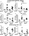
IRF-3 supports expression of select interferon-stimulated genes (ISG) but not the generation of MHV68-specific CD8 T cells during the peak of MHV68 latent infection. Mice were infected as for Fig. 1. At 16 days postinfection, total RNA was isolated from individual spleens and subjected to qRT-PCR using CXCL9 (A), MX2 (B), and MNDA (C) primers, with relative expression further normalized against corresponding glyceraldehyde-3-phosphate dehydrogenase (GAPDH) levels and relative to expression observed in mock-infected BL6 mice. (D) Sera from mock- or MHV68-infected mice were subjected to IFN-γ ELISA. Frequency (E) and absolute number (F) of MHV68-positive CD8 T cells (defined as CD3+ CD8+ CD44+ orf6 tetramer+) were quantified in the spleens harvested at 16 days postinfection. Each symbol represents an individual mouse; data were pooled from 2 to 3 independent experiments. *, P < 0.05.
FIG 5
FIG 5
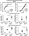
IRF-3 expression is dispensable for the establishment of MHV68 splenic latency following intraperitoneal infection. Mice of indicated genotypes were intraperitoneally infected with 1,000 PFU of wild-type MHV68 and analyzed at 16 days postinfection. Splenocytes were pooled from mice within each group and subjected to limiting dilution analyses to define the frequency of MHV68 DNA-positive cells (A) and ex vivo reactivation (B). Germinal center B cells (C and D) and T follicular helper cells (E and F) were defined as for Fig. 2 and measured in splenocytes from individual mice (each symbol represents individual animal). Data were pooled from 2 individual experiments. *, P < 0.05.
FIG 6
FIG 6
IRF-3 deficiency does not rescue attenuated acute replication of the gammaherpesvirus protein kinase null mutant. BL6 and IRF-3−/− mice were intranasally infected with 10,000 PFU of N36S MHV68 mutant that lacks expression of viral protein kinase or a corresponding control MHV68. (A) Viral titers in the lungs were determined at 7 days postinfection. Dotted line represents the limit of infectious virus detection. (B to D) Levels of mRNA of indicated ISGs were measured in total RNA isolated from lungs using qRT-PCR. Relative expression was normalized to corresponding GAPDH levels and to the relative expression observed in mock-infected BL6 mice. Each symbol represents an individual mouse. *, P < 0.05.
FIG 7
FIG 7
IRF-3 deficiency does not rescue attenuated latency of the gammaherpesvirus protein kinase null mutant. BL6 and IRF-3−/− mice were intranasally infected with 10,000 PFU of N36S MHV68 mutant that lacks expression of viral protein kinase or a corresponding control MHV68. At 16 days postinfection, frequencies of MHV68 DNA-positive cells (A) and viral reactivation (B) were assessed in splenocytes pooled from 3 to 4 mice/group as for Fig. 1A and B. Data were pooled from 4 to 5 independent experiments. (C to E) Levels of indicated mRNAs were determined by qRT-PCR in spleens and were further normalized to corresponding GAPDH mRNA levels, with fold changes defined on the basis of the relative expression in spleens of mock-infected BL6 mice. Each symbol represents an individual spleen. *, P < 0.05.
FIG 8
FIG 8
IRF-3 deficiency partially rescues induction of CD4 T follicular helper cells and class-switched plasma cells but not germinal center B cells in N36S-infected mice. BL6 and IRF-3−/− mice were infected with 10,000 PFU of wild-type (WT) or N36S MHV68, as for Fig. 6, with splenocytes analyzed at 16 days postinfection using the gating strategy shown in Fig. 2. Germinal center B cells were identified as GL7+ CD95+ cells pregated on B220+ cells and expressed as frequency (A) and total cell number (B). T follicular helper cells were identified as PD1+ CXCR5+ cells pregated on CD3+ CD4+ cells and expressed as frequency (C) and total cell number (D). Plasma cells were identified as IgD-IRF4+ GL7 cells pregated on B220int/+ GL7 cells expressed as frequency (E) and total cell number (F). Each symbol represents the result for an individual spleen, and data from 2 to 4 independent experiments were pooled. *, P < 0.05.

Similar articles

Cited by

References

    1. Au WC, Moore PA, Lowther W, Juang YT, Pitha PM. 1995. Identification of a member of the interferon regulatory factor family that binds to the interferon-stimulated response element and activates expression of interferon-induced genes. Proc Natl Acad Sci U S A 92:11657–11661. doi:10.1073/pnas.92.25.11657. - DOI - PMC - PubMed
    1. Sato M, Suemori H, Hata N, Asagiri M, Ogasawara K, Nakao K, Nakaya T, Katsuki M, Noguchi S, Tanaka N, Taniguchi T. 2000. Distinct and essential roles of transcription factors IRF-3 and IRF-7 in response to viruses for IFN-alpha/beta gene induction. Immunity 13:539–548. doi:10.1016/s1074-7613(00)00053-4. - DOI - PubMed
    1. Honda K, Takaoka A, Taniguchi T. 2006. Type I interferon gene induction by the interferon regulatory factor family of transcription factors. Immunity 25:349–360. doi:10.1016/j.immuni.2006.08.009. - DOI - PubMed
    1. Menachery VD, Leib DA. 2009. Control of herpes simplex virus replication is mediated through an interferon regulatory factor 3-dependent pathway. J Virol 83:12399–12406. doi:10.1128/JVI.00888-09. - DOI - PMC - PubMed
    1. Thackray LB, Duan E, Lazear HM, Kambal A, Schreiber RD, Diamond MS, Virgin HW. 2012. Critical role for interferon regulatory factor 3 (IRF-3) and IRF-7 in type I interferon-mediated control of murine norovirus replication. J Virol 86:13515–13523. doi:10.1128/JVI.01824-12. - DOI - PMC - PubMed

Publication types

Substances

LinkOut - more resources