. 2021 Mar 26;371(6536):1379-1382.
doi: 10.1126/science.abf4896. Epub 2021 Feb 17.

Intranasal fusion inhibitory lipopeptide prevents direct-contact SARS-CoV-2 transmission in ferrets

Affiliations

Intranasal fusion inhibitory lipopeptide prevents direct-contact SARS-CoV-2 transmission in ferrets

Rory D de Vries et al. Science. .

Abstract

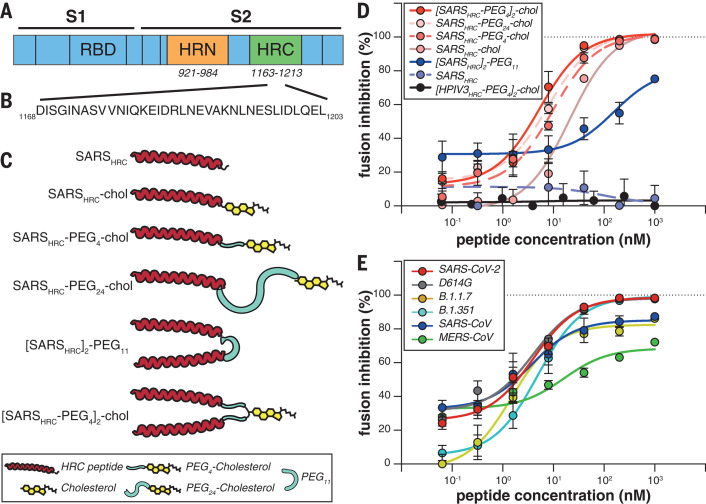
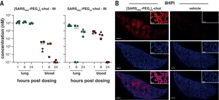
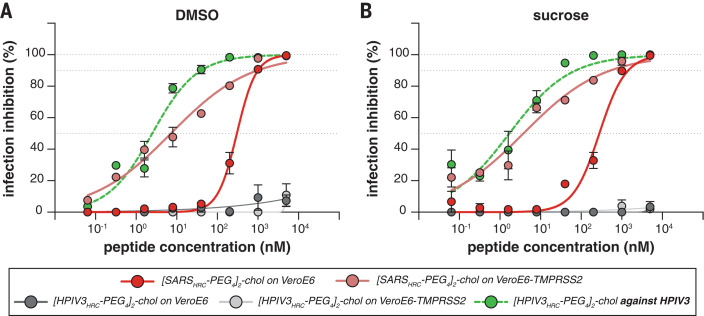
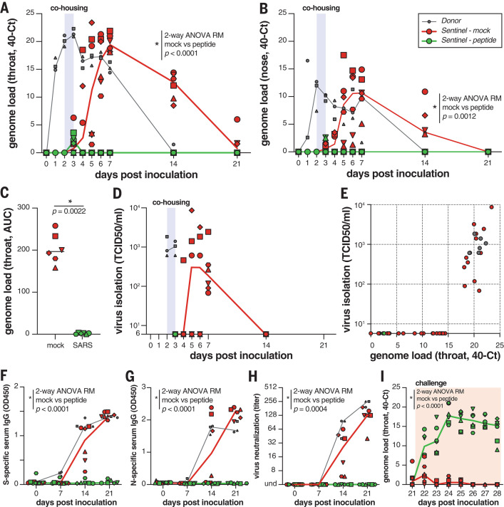
Containment of the COVID-19 pandemic requires reducing viral transmission. Severe acute respiratory syndrome coronavirus 2 (SARS-CoV-2) infection is initiated by membrane fusion between the viral and host cell membranes, which is mediated by the viral spike protein. We have designed lipopeptide fusion inhibitors that block this critical first step of infection and, on the basis of in vitro efficacy and in vivo biodistribution, selected a dimeric form for evaluation in an animal model. Daily intranasal administration to ferrets completely prevented SARS-CoV-2 direct-contact transmission during 24-hour cohousing with infected animals, under stringent conditions that resulted in infection of 100% of untreated animals. These lipopeptides are highly stable and thus may readily translate into safe and effective intranasal prophylaxis to reduce transmission of SARS-CoV-2.


Figures

Fig. 1
Fig. 1. Peptide-lipid conjugates that inhibit SARS-CoV-2 spike (S)mediated fusion.
(A) The functional domains of SARS-CoV-2 S protein, the receptor binding domain (RBD) and heptad repeats (HRN and HRC), are indicated. (B) Sequence of the peptides derived from the HRC domain of SARS-CoV-2 S. Single-letter abbreviations for the amino acid residues are as follows: A, Ala; C, Cys; D, Asp; E, Glu; F, Phe; G, Gly; H, His; I, Ile; K, Lys; L, Leu; M, Met; N, Asn; P, Pro; Q, Gln; R, Arg; S, Ser; T, Thr; V, Val; W, Trp; and Y, Tyr. (C) Monomeric and dimeric forms of lipid-tagged SARS-CoV-2 inhibitory peptides that were assessed in cellcell fusion assays. (D) Cellcell fusion assays with different inhibitory peptides. The percentage inhibition is shown for six different SARS-CoV-2specific peptides and a control HPIV-3specific peptide at increasing concentrations. Percent inhibition was calculated as the ratio of the relative luminescence units in the presence of a specific concentration of inhibitor (X) and the relative luminescence units in the absence of inhibitor, corrected for background luminescence. Percent inhibition = 100 [1 (luminescence at X background)/(luminescence in absence of inhibitor background)]. The difference between the results for [SARSHRC-PEG4]2-chol and SARSHRC-PEG4-chol lipopeptides was statistically significant [two-way analysis of variance (ANOVA), P < 0.0001]. (E) Fusion inhibitory activity of [SARSHRC-PEG4]2-chol peptide against emerging SARS-CoV-2 S variants, MERS-CoV-2 S, and SARS-CoV S. Data in (D) and (E) are means standard error of the mean (SEM) from three separate experiments, with the curve representing a four-parameter dose-response model.
Fig. 2
Fig. 2. Biodistribution of [SARSHRC-PEG4]2-chol and SARSHRC-PEG24 peptides after intranasal administration to mice.
(A) The concentration of lipopeptides (y axis) was measured by ELISA in lung homogenates and plasma samples (n = 4 mice, with the exception of [SARSHRC-PEG4]2-chol IN, for which n = 3 at 8 and 24 hours, and n = 1 for vehicle treatment). Median is indicated by horizontal bar. (B) Lung sections of [SARSHRC-PEG4]2-chol-treated (or vehicle-treated) mice were stained with anti-SARS-HRC antibody (red) confirming broad distribution of [SARSHRC-PEG4]2-chol in the lung (8 hours post inoculation, 8HPI). Scale bar, 500 m in lung tile scan and 50 m in magnification; representative images and a full tile scan are shown. Nuclei were counterstained with 4,6-diamidino-2-phenylindole (blue).
Fig. 3
Fig. 3. Inhibition of infectious SARS-CoV-2 entry by [SARSHRC-PEG4]2-chol and [HPIV-3HRC-PEG4]2-chol peptides.
(A and B) The percentage inhibition of infection is shown on VeroE6 and VeroE6-TMPRSS2 cells with increasing concentrations of [SARSHRC-PEG4]2-chol (red lines) and [HPIV-3HRC-PEG4]2-chol (gray lines). DMSO-formulated (A) and sucrose-formulated (B) stocks were tested side by side. Mean SEM of triplicates is shown; dotted lines show 50% and 90% inhibition. Additionally, the potency of [HPIV-3HRC-PEG4]2-chol was confirmed by inhibition of infectious HPIV-3 entry (dotted green lines, performed on Vero cells).
Fig. 4
Fig. 4. [SARSHRC-PEG4]2-chol prevents SARS-CoV-2 transmission in vivo.
(A and B) Viral loads detected in throat (A) and nose (B) swabs by RT-qPCR. (C) Comparison of the AUC from genome loads reported in (B) for mock- and peptide-treated sentinels. (D) Viral loads detected in throat swabs by virus isolation on VeroE6. (E) Correlation between viral loads in the throat as detected via RT-qPCR and virus isolation. Presence of anti-S (F) or anti-N (G) antibodies was determined by IgG ELISA assay. Presence of neutralizing antibodies was determined in a virus neutralization assay (H). Virus neutralizing antibodies are displayed as the end-point serum dilution factor that blocks SARS-CoV-2 replication. Direct inoculation of peptide-treated or mock-treated animals with SARS-CoV-2 led to productive infection in only the previously peptide-treated animals (I), in the absence of S-specific, N-specific, and neutralizing antibodies. Donor animals shown in gray, mock-treated animals in red, peptide-treated animals in green. Symbols correspond to individual animals (defined in fig. S6). Line graphs in (A), (B), (D), and (F) to (I) connect the median of individual animals per time point. Mock- and peptide-treated groups were compared using two-way ANOVA repeated measures [(A), (B), and (F) to (I)] or Mann-Whitney test (C).

Update of

Similar articles

Cited by

References

    1. Li F., Structure, function, and evolution of coronavirus spike proteins. Annu. Rev. Virol. 3, 237–261. (2016). 10.1146/annurev-virology-110615-042301 - DOI - PMC - PubMed
    1. Hoffmann M., Kleine-Weber H., Schroeder S., Krger N., Herrler T., Erichsen S., Schiergens T. S., Herrler G., Wu N.-H., Nitsche A., Mller M. A., Drosten C., Phlmann S., SARS-CoV-2 cell entry depends on ACE2 and TMPRSS2 and is blocked by a clinically proven protease inhibitor. Cell 181, 271–280.e8. (2020). 10.1016/j.cell.2020.02.052 - DOI - PMC - PubMed
    1. Wan Y., Shang J., Graham R., Baric R. S., Li F., Receptor recognition by the novel coronavirus from Wuhan: an analysis based on decade-long structural studies of SARS coronavirus. J. Virol. 94, e00127-20 (2020). 10.1128/JVI.00127-20 - DOI - PMC - PubMed
    1. Outlaw V. K., Bovier F. T., Mears M. C., Cajimat M. N., Zhu Y., Lin M. J., Addetia A., Lieberman N. A. P., Peddu V., Xie X., Shi P.-Y., Greninger A. L., Gellman S. H., Bente D. A., Moscona A., Porotto M., Inhibition of coronavirus entry in vitro and ex vivo by a lipid-conjugated peptide derived from the SARS-CoV-2 spike glycoprotein HRC domain. mBio 11, e01935-20 (2020). 10.1128/mBio.01935-20 - DOI - PMC - PubMed
    1. Xia S., Yan L., Xu W., Agrawal A. S., Algaissi A., Tseng C. K., Wang Q., Du L., Tan W., Wilson I. A., Jiang S., Yang B., Lu L., A pan-coronavirus fusion inhibitor targeting the HR1 domain of human coronavirus spike. Sci. Adv. 5, eaav4580 (2019). 10.1126/sciadv.aav4580 - DOI - PMC - PubMed

Publication types

MeSH terms

Substances