. 2021 Feb 17;7(8):eabf1738.
doi: 10.1126/sciadv.abf1738. Print 2021 Feb.

An engineered decoy receptor for SARS-CoV-2 broadly binds protein S sequence variants

Affiliations

An engineered decoy receptor for SARS-CoV-2 broadly binds protein S sequence variants

Kui K Chan et al. Sci Adv. .

Abstract

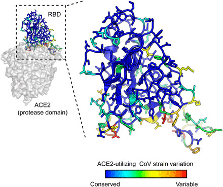
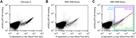
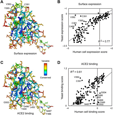
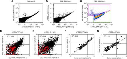
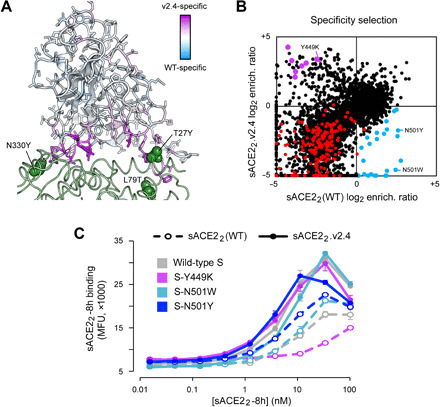
The spike S of SARS-CoV-2 recognizes ACE2 on the host cell membrane to initiate entry. Soluble decoy receptors, in which the ACE2 ectodomain is engineered to block S with high affinity, potently neutralize infection and, because of close similarity with the natural receptor, hold out the promise of being broadly active against virus variants without opportunity for escape. Here, we directly test this hypothesis. We find that an engineered decoy receptor, sACE22v2.4, tightly binds S of SARS-associated viruses from humans and bats, despite the ACE2-binding surface being a region of high diversity. Saturation mutagenesis of the receptor-binding domain followed by in vitro selection, with wild-type ACE2 and the engineered decoy competing for binding sites, failed to find S mutants that discriminate in favor of the wild-type receptor. We conclude that resistance to engineered decoys will be rare and that decoys may be active against future outbreaks of SARS-associated betacoronaviruses.


Figures

Fig. 1
Fig. 1. SARS-associated coronaviruses have high sequence diversity at the ACE2-binding site.
The RBD of SARS-CoV-2 [Protein Data Bank (PDB): 6M17] is colored by diversity between seven SARS-associated CoV strains (blue, conserved; red, variable).
Fig. 2
Fig. 2. FACS selection for variants of S with high or low binding signal to ACE2.
(A) Flow cytometry analysis of Expi293F cells expressing full-length S of SARS-CoV-2 with an N-terminal c-myc tag. Staining for the myc-epitope is on the x axis, while the detection of bound sACE22-8h (2.5 nM) is on the y axis. S plasmid was diluted 1500-fold by weight with carrier DNA so that cells typically express no more than one coding variant; under these conditions, most cells are negative. (B) Flow cytometry of cells transfected with the RBD single site-saturation mutagenesis (SSM) library shows cells expressing S variants with reduced sACE22-8h binding. (C) Gating strategy for FACS. S-expressing cells positive for the c-myc epitope were gated (blue), and the highest (“ACE2-high”) and lowest (“ACE2-low”) 20% of cells with bound sACE22-8h relative to myc-S expression were collected.
Fig. 3
Fig. 3. Deep mutagenesis reveals that the ACE2-binding site of SARS-CoV-2 tolerates many mutations.
(A) Positional scores for surface expression are mapped to the structure of the SARS-CoV-2 RBD (PDB: 6M17, oriented as in Fig. 1). Blue residues in the protein core are highly conserved in the FACS selection for surface S expression (judged by depletion of mutations from the ACE2-high and ACE2-low gates), while surface residues in red tolerate mutations. (B) Correlation plot of expression scores from mutant selection in human cells of full-length S (x axis) versus the conservation scores (mean of the log2 enrichment ratios at a residue position) from mutant selection in the isolated RBD by yeast display (y axis). Notable outliers are indicated. (C) Conservation scores from the ACE2-high gated cell population are mapped to the RBD structure, with residues colored from low (blue) to high (red) mutational tolerance. (D) Correlation plot of RBD conservation scores for high ACE2 binding from deep mutagenesis of S in human cells (x axis) versus deep mutagenesis of the RBD on the yeast surface (mean of ΔKD app; y axis).
Fig. 4
Fig. 4. A competition-based selection to identify RBD mutations within S of SARS-CoV-2 that preferentially bind WT or engineered ACE2 receptors.
(A) Expi293F cells were transfected with WT myc-S and incubated with competing sACE22(WT)-IgG1 (25 nM) and sACE22.v2.4-8h (20 nM). Bound protein was detected by flow cytometry after immunostaining for the respective epitope tags. (B) As in (A), except cells were transfected with the RBD SSM library. A population of cells expressing S variants with increased specificity toward sACE22.v2.4 is apparent (cells shifted to the upper left of the main population). (C) Gates used for FACS of cells expressing the RBD SSM library. After excluding cells without bound protein, the top 20% of cells for bound sACE22.v2.4-8h (magenta gate) and for bound sACE22(WT)-IgG1 (green gate) were collected. (D and E) Agreement between log2 enrichment ratios from two independent FACS selections for cells expressing S variants with increased specificity for (D) sACE22(WT) or (E) sACE22.v2.4. R2 values are calculated for nonsynonymous mutations (black). Nonsense mutations are red. (F and G) Conservation scores are calculated from the mean of the log2 enrichment ratios for all nonsynonymous substitutions at a given residue position. Correlation plots show agreement between conservation scores for two independent selections for cells within the (D) sACE22(WT)- or (E) sACE22.v2.4-specific gates.
Fig. 5
Fig. 5. Mutations within the RBD that confer specificity toward WT ACE2 are rare.
(A) The SARS-CoV-2 RBD is colored by specificity score [the difference between the conservation scores for cells collected in the sACE22(WT)- and sACE22.v2.4-specific gates]. Residues that are hotspots for mutations with increased specificity toward sACE22(WT) are blue or toward sACE22.v2.4 are purple. The contacting surface of ACE2 is shown as a green ribbon, with sites of mutations in sACE22.v2.4 labeled and shown as green spheres. (B) Log2 enrichment ratios for mutations in S expressed by cell populations collected in the sACE22(WT)-specific (x axis) and sACE22.v2.4-specific (y axis) gates. Data are the mean from two independent sorting experiments. S mutants in blue were predicted to have increased specificity for sACE22(WT) and were tested by targeted mutagenesis in fig. S6. S mutants in purple were predicted to have increased specificity for sACE22.v2.4 and were tested by targeted mutagenesis in fig. S7. Other nonsynonymous mutations are black. Nonsense mutations are red. (C) WT myc-S (gray) and three variants, Y449K (purple), N501W (light blue), and N501Y (dark blue), were expressed in Expi293F cells and tested by flow cytometry for binding to sACE22(WT)-8h (dashed lines) or sACE22.v2.4-8h (solid lines). MFU, mean fluorescence units.

Update of

References

    1. Zhou P., Yang X.-L., Wang X.-G., Hu B., Zhang L., Zhang W., Si H.-R., Zhu Y., Li B., Huang C.-L., Chen H.-D., Chen J., Luo Y., Guo H., Jiang R.-D., Liu M.-Q., Chen Y., Shen X.-R., Wang X., Zheng X.-S., Zhao K., Chen Q.-J., Deng F., Liu L.-L., Yan B., Zhan F.-X., Wang Y.-Y., Xiao G.-F., Shi Z.-L., A pneumonia outbreak associated with a new coronavirus of probable bat origin. Nature 579, 270–273 (2020). - PMC - PubMed
    1. Walls A. C., Park Y.-J., Tortorici M. A., Wall A., McGuire A. T., Veesler D., Structure, function, and antigenicity of the SARS-CoV-2 spike glycoprotein. Cell 181, 281–292.e6 (2020). - PMC - PubMed
    1. Wan Y., Shang J., Graham R., Baric R. S., Li F., Receptor recognition by novel coronavirus from Wuhan: An analysis based on decade-long structural studies of SARS coronavirus. J. Virol. 94, e00127-20 (2020). - PMC - PubMed
    1. Wrapp D., Wang N., Corbett K. S., Goldsmith J. A., Hsieh C.-L., Abiona O., Graham B. S., McLellan J. S., Cryo-EM structure of the 2019-nCoV spike in the prefusion conformation. Science 367, 1260–1263 (2020). - PMC - PubMed
    1. Hoffmann M., Kleine-Weber H., Schroeder S., Krüger N., Herrler T., Erichsen S., Schiergens T. S., Herrler G., Wu N.-H., Nitsche A., Müller M. A., Drosten C., Pöhlmann S., SARS-CoV-2 cell entry depends on ACE2 and TMPRSS2 and is blocked by a clinically proven protease inhibitor. Cell 181, 271–280.e8 (2020). - PMC - PubMed

Publication types

Substances