. 2021 Feb 23;118(8):e2019152118.
doi: 10.1073/pnas.2019152118.

SOX9 keeps growth plates and articular cartilage healthy by inhibiting chondrocyte dedifferentiation/osteoblastic redifferentiation

Affiliations

SOX9 keeps growth plates and articular cartilage healthy by inhibiting chondrocyte dedifferentiation/osteoblastic redifferentiation

Abdul Haseeb et al. Proc Natl Acad Sci U S A. .

Abstract

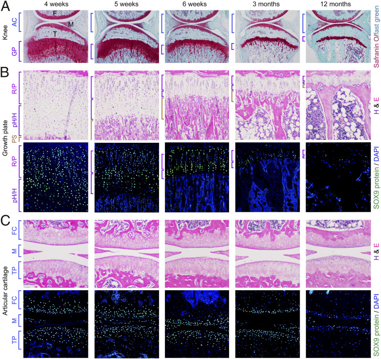
Cartilage is essential throughout vertebrate life. It starts developing in embryos when osteochondroprogenitor cells commit to chondrogenesis, activate a pancartilaginous program to form cartilaginous skeletal primordia, and also embrace a growth-plate program to drive skeletal growth or an articular program to build permanent joint cartilage. Various forms of cartilage malformation and degeneration diseases afflict humans, but underlying mechanisms are still incompletely understood and treatment options suboptimal. The transcription factor SOX9 is required for embryonic chondrogenesis, but its postnatal roles remain unclear, despite evidence that it is down-regulated in osteoarthritis and heterozygously inactivated in campomelic dysplasia, a severe skeletal dysplasia characterized postnatally by small stature and kyphoscoliosis. Using conditional knockout mice and high-throughput sequencing assays, we show here that SOX9 is required postnatally to prevent growth-plate closure and preosteoarthritic deterioration of articular cartilage. Its deficiency prompts growth-plate chondrocytes at all stages to swiftly reach a terminal/dedifferentiated stage marked by expression of chondrocyte-specific (Mgp) and progenitor-specific (Nt5e and Sox4) genes. Up-regulation of osteogenic genes (Runx2, Sp7, and Postn) and overt osteoblastogenesis quickly ensue. SOX9 deficiency does not perturb the articular program, except in load-bearing regions, where it also provokes chondrocyte-to-osteoblast conversion via a progenitor stage. Pathway analyses support roles for SOX9 in controlling TGFβ and BMP signaling activities during this cell lineage transition. Altogether, these findings deepen our current understanding of the cellular and molecular mechanisms that specifically ensure lifelong growth-plate and articular cartilage vigor by identifying osteogenic plasticity of growth-plate and articular chondrocytes and a SOX9-countered chondrocyte dedifferentiation/osteoblast redifferentiation process.

Keywords: SOX9; cartilage; cell differentiation; lineage determination; transcriptional regulation.


Conflict of interest statement

The authors declare no competing interest.

Figures

Fig. 1.
Fig. 1.
SOX9 expression declines as GPs retire and AC ages. (A) Sections through the knee of 4-wk- to 12-mo-old mice. Staining, Safranin O (cartilage) and Fast Green. F, femur; M, meniscus; T, tibia. (B) Tibia proximal GPs. (Top) Hematoxylin and eosin (H&E) staining. (Bottom) SOX9 antibody (green) and DAPI (blue, cell nuclei) staining. R/P, relesting and proliferative zones; pH/H, prehypertrophic and hypertrophic zones. PS, primary spongiosa. (C) Knee joint. FC, femoral condyle AC; M, menisci; TP, tibial plateau AC.
Fig. 2.
Fig. 2.
SOX9 is required to keep GPs open and AC healthy. (A) Tibia proximal GPs and knee sections from control and SOX9 mutant mice treated and analyzed at various time points as indicated. Staining, Safranin O/Fast Green. Note that, in this and all other figures showing several time points, control pictures are displayed at one time point only, but these pictures are representative of the data obtained at all time points. (B, Left) Sections through medial femoral condyles (MFCs) and tibial plateaus (MTPs) of control and mutant mice treated as indicated. Staining, Safranin O/Fast Green. (B, Right) OARSI scores. Dots, scores for individual mice. Bars and brackets, means ± SD for six to eight animals. P values are indicated for statistical significance of differences between controls and mutants calculated using Student’s t test.
Fig. 3.
Fig. 3.
SOX9 ensures high-level expression of PC and GP markers. (A) RNA-seq assay. Three pairs of control and mutant mice received tamoxifen at 3 mo and were analyzed 7 or 14 d later, as indicated. Averages of NRPKM values obtained for control and mutant samples, and statistically significant (P < 0.05) differences obtained in paired t tests for control and mutant data are indicated. (B) In situ detection of Acan, Ihh, and Col10a1 RNA (magenta) in tibia GP and knee AC sections. Sox9 control and mutant mice were given tamoxifen at 3 mo and analyzed at indicated times. Ihh RNA signals were amplified with Adobe Photoshop. Arrows, cells positive for Ihh RNA. Counterstaining, hematoxylin. See SI Appendix, Fig. S4E, for entire-knee pictures.
Fig. 4.
Fig. 4.
SOX9 is dispensable for AC marker expression, except in load-bearing regions. (A) RNA-seq assay of AC markers. Data were generated and plotted as in Fig. 3A. (B) In situ detection of Prg4, Smoc2, and Clu RNAs (magenta). Sox9 control and mutant mice were given tamoxifen at 3 mo and analyzed 15 d later. Counterstaining, hematoxylin. Green arrows, Smoc2- and Clu-expressing GPCs. Black arrows, load-bearing AC regions down-regulating AC markers. (C) Same experiment as in B, except that mice were analyzed 5 mo after tamoxifen treatment. (D) RNA-seq assay of early-osteoarthritis and inflammation markers. Data were generated and plotted as in Fig. 3A. P values are indicated for differences between control and mutant samples reaching (P < 0.05) and approaching (P < 0.25) statistical significance.
Fig. 5.
Fig. 5.
Sox9 inactivation prompts chondrocyte osteoblastogenesis. (A) RNA-seq assay of OB markers. Data were generated and plotted as in Figs. 3A and 4D. (B) In situ detection of Runx2, Sp7, and Bglap/Bglap2 RNAs (magenta) in tibia proximal GP and knee AC. Sox9 control and mutant mice received tamoxifen at 3 mo and were analyzed at indicated times. Counterstaining, hematoxylin. RNA signals for Runx2 and Sp7 were amplified using Adobe Photoshop. See SI Appendix, Fig. S5A, for entire-knee sections. (C) In situ detection of tdTomato (red) and RUNX2 and osteocalcin immunostaining (green). Sox9 control and mutant mice received tamoxifen at 3 mo and were analyzed at indicated times. Cell nuclei are stained with DAPI. Blue brackets, GP. Note the progressive disappearance of RUNX2 signal in the mutant GP 2 and 4 d after tamoxifen treatment, reflecting the loss of hypertrophic GPCs and the fact that nonhypertrophic GPCs have not up-regulated RUNX2 expression yet.
Fig. 6.
Fig. 6.
SOX9 prevents chondrocyte dedifferentiation/osteoblastic redifferentiation. (A) UMAP plot of skeletal cell populations extracted from the femoral and tibial epiphyses of two pairs of control (Sox9fl/fl) and mutant (Sox9fl/flAcanCreERT2/+) mouse littermates. Mice were treated with tamoxifen 4 and 5 d before analysis at P13 and P19. Cells were segregated into 35 clusters, as indicated. (B) Dot plot showing the relative expression of marker genes across clusters. The darkness and size of the dots reflect the average RNA level in expressing cells and the proportion of expressing cells, respectively. (C) Monocle pseudotemporal ordering of cell clusters. (D) RNA in situ detection of terminal GPC and skeletal progenitor markers (magenta) in Sox9 control and mutant mice treated with tamoxifen at 3 mo and analyzed 4 to 14 d later. The Nt5e and Sox4 RNA signals were amplified using Adobe Photoshop. See SI Appendix, Fig. S8A, for entire-knee sections.
Fig. 7.
Fig. 7.
TGFβ and BMP signaling likely contributes to chondrocyte fate changes upon Sox9 inactivation. (A) pSMAD2/3 immunostaining in Sox9 control and mutant mice given tamoxifen at 3 mo and analyzed at indicated times. Cell nuclei are stained with DAPI. Blue brackets, GPs. Graphs, pSMAD2/3 signal intensities. Dots, values obtained for individual mice. Bars and brackets, means ± SD for four animals per time point. P values obtained in t tests of differences between controls and mutants in the first week (days 2 to 7 combined), second and third week (days 10 to 21 combined), and 2 and 9 mo after Sox9 inactivation are shown in purple. P values for differences between early (2 to 7 d) and late (10 to 21 d) times are shown in blue for controls and in red for mutants. (B) pSMAD1/5/9 immunostaining conducted and presented as in A, except that differences between controls and mutants were assessed at 2 to 4 d and 7 to 21 d after Sox9 inactivation. (C) RNA levels of various genes in Sox9fl/fl primary chondrocytes transduced with Ad-GFP (controls) or Ad-CRE (mutant) adenovirus, and treated with 5 ng/mL TGFβ1 or 200 ng/mL BMP2 for 24 h. RNA values were measured by qRT-PCR and calculated relative to those for Hprt and control cells (Ad-GFP, no additive). Dots, individual values obtained in four independent experiments, each with one pair of control and mutant cells. Average values and SDs are shown. P values of ≤0.05 in paired t tests are indicated for conditions testing the effects of Sox9 inactivation and/or the effects of growth factors. (D) Western blots of β-actin, SOX9, RUNX2, and SP7 present in extracts from cells treated as in C. The RUNX2/β-actin and SP7/β-actin ratios are presented relative to that obtained for cells treated with Ad-GFP, and with Ad-GFP/BMP2, respectively. Data are representative of multiple similar experiments.
Fig. 8.
Fig. 8.
Roles of SOX9 in established GPs and AC. (A) SOX9 is required to keep GPs open and prevent AC from preosteoarthritic degeneration, namely by preventing osteoblastogenesis of GPCs and load-bearing ACCs. (B) SOX9 ensures and paces the multistep differentiation of GPCs and helps maintain the ACC permanent phenotype, at least in part by preventing GPCs and ACCs from reaching terminal and progenitor stages leading to osteoblast differentiation.

Comment in

  • Keep your Sox on, chondrocytes!
    Pitsillides AA, Beier F. Pitsillides AA, et al. Nat Rev Rheumatol. 2021 Jul;17(7):383-384. doi: 10.1038/s41584-021-00628-9. Nat Rev Rheumatol. 2021. PMID: 33953371 No abstract available.

References

    1. Merrell A. J., Stanger B. Z., Adult cell plasticity in vivo: De-differentiation and transdifferentiation are back in style. Nat. Rev. Mol. Cell Biol. 17, 413–425 (2016). - PMC - PubMed
    1. Gupta P. B., Pastushenko I., Skibinski A., Blanpain C., Kuperwasser C., Phenotypic plasticity: Driver of cancer initiation, progression, and therapy resistance. Cell Stem Cell 24, 65–78 (2019). - PMC - PubMed
    1. Kaji D. A., et al. ., Cellular plasticity in musculoskeletal development, regeneration, and disease. J. Orthop. Res. 38, 708–718 (2020). - PMC - PubMed
    1. Piera-Velazquez S., Jimenez S. A., Endothelial to mesenchymal transition: Role in physiology and in the pathogenesis of human diseases. Physiol. Rev. 99, 1281–1324 (2019). - PMC - PubMed
    1. Hallett S. A., Ono W., Ono N., Growth plate chondrocytes: Skeletal development, growth and beyond. Int. J. Mol. Sci. 20, E6009 (2019). - PMC - PubMed

Publication types

Substances

LinkOut - more resources