. 2021 Feb 23;118(8):e2024512118.
doi: 10.1073/pnas.2024512118.

Molecular mechanisms of assembly and TRIP13-mediated remodeling of the human Shieldin complex

Affiliations

Molecular mechanisms of assembly and TRIP13-mediated remodeling of the human Shieldin complex

Wei Xie et al. Proc Natl Acad Sci U S A. .

Abstract

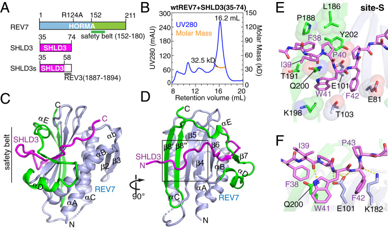
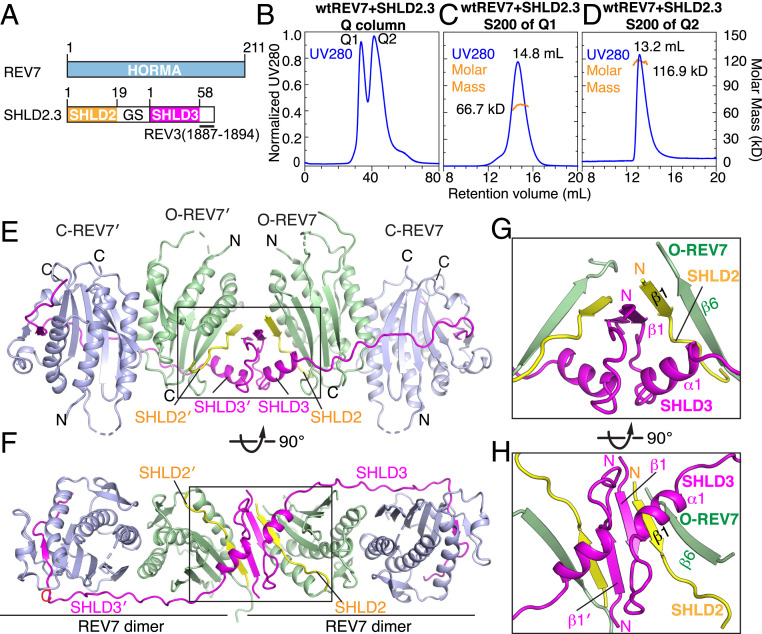
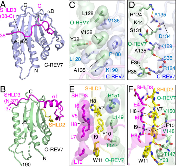
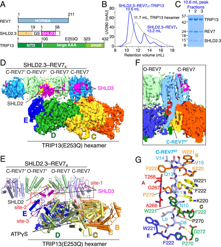
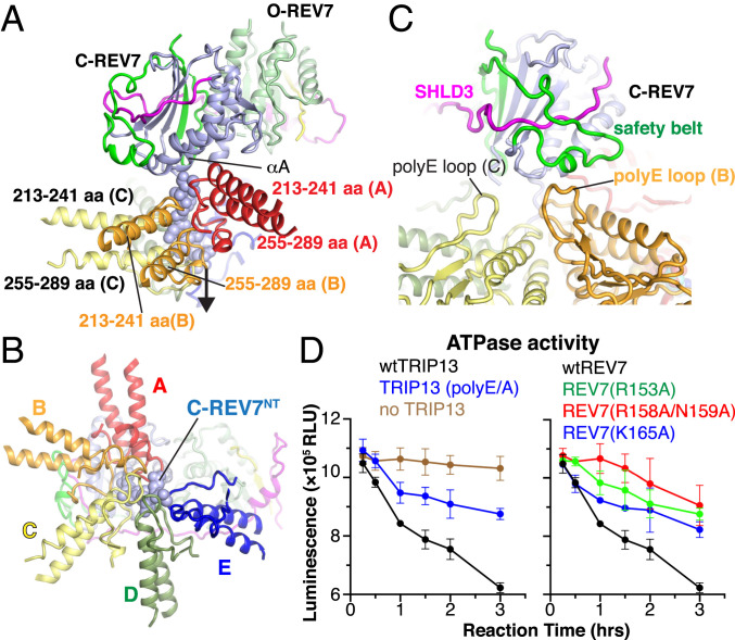
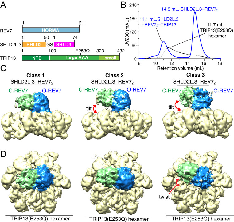
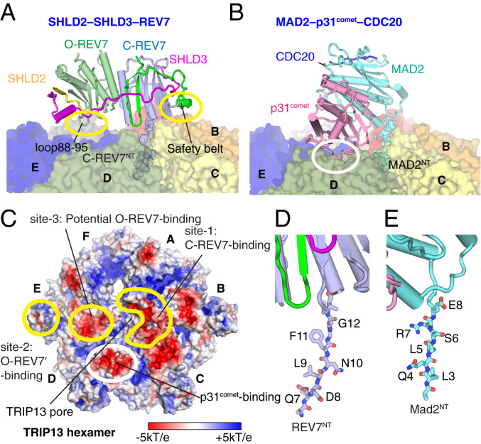
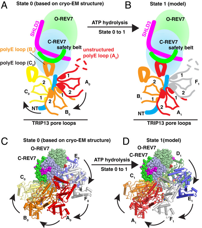
The Shieldin complex, composed of REV7, SHLD1, SHLD2, and SHLD3, protects DNA double-strand breaks (DSBs) to promote nonhomologous end joining. The AAA+ ATPase TRIP13 remodels Shieldin to regulate DNA repair pathway choice. Here we report crystal structures of human SHLD3-REV7 binary and fused SHLD2-SHLD3-REV7 ternary complexes, revealing that assembly of Shieldin requires fused SHLD2-SHLD3 induced conformational heterodimerization of open (O-REV7) and closed (C-REV7) forms of REV7. We also report the cryogenic electron microscopy (cryo-EM) structures of the ATPγS-bound fused SHLD2-SHLD3-REV7-TRIP13 complexes, uncovering the principles underlying the TRIP13-mediated disassembly mechanism of the Shieldin complex. We demonstrate that the N terminus of REV7 inserts into the central channel of TRIP13, setting the stage for pulling the unfolded N-terminal peptide of C-REV7 through the central TRIP13 hexameric channel. The primary interface involves contacts between the safety-belt segment of C-REV7 and a conserved and negatively charged loop of TRIP13. This process is mediated by ATP hydrolysis-triggered rotatory motions of the TRIP13 ATPase, thereby resulting in the disassembly of the Shieldin complex.

Keywords: SHLD2–SHLD3–REV7 complex; SHLD2–SHLD3–REV7–TRIP13 complex; SHLD3–REV7 complex; Shieldin assembly; TRIP13-mediated disassembly of Shieldin.


Conflict of interest statement

The authors declare no competing interest.

Figures

Fig. 1.
Fig. 1.
Crystal structure of SHLD3s–REV7 monomer complex reveals safety-belt topology and site-S interface. (A) Schematic drawing of human REV7 and SHLD3s fusion protein. The safety-belt segment of REV7 spans residues 152 to 180. (B) Purification of the SHLD3 (35 to 74)–REV7 monomer complex on an S200 gel filtration column. The major peak exhibited a mol. wt. = 32.5 kDa by SEC-MALS. (C and D) Two views of the overall structure of the SHLD3s–REV7 monomer complex. The N- and C-terminal halves of REV7 monomer are colored in light blue and green, respectively, while SHLD3s is colored in magenta. A black box highlights the site-S region in D. (E and F) Hydrophobic (E) and hydrogen bonding (F) interactions involving site-S. As shown in E, the bulky side chain of SHLD3 Phe-38 wedges into the hydrophobic pocket lined by REV7 residues Leu-186, Pro-188, Thr-191, and Tyr-202, while SHLD3 Trp-41 and Phe-42 stack tightly with the side chains of Glu-81, Glu-101, Thr-103, Lys-198, and Gln-200 of REV7. As shown in F, the backbones of SHLD3 Ile-39, Trp-41, Phe-42, and Pro-43 further interacts with the side chains of REV7 residues Glu-101, Lys-182, and Leu-173, and Ala-174 by hydrogen bonding.
Fig. 2.
Fig. 2.
Crystal structure of SHLD2.3–REV74 complex. (A) Schematic drawing of human REV7 and SHLD2.3 fusion protein. (B) Q column purification of the complex SHLD2.3 bound to REV7 yields two peaks labeled Q1 and Q2. (C) Size exclusion S200 purification of peak labeled Q1 and measurement of a 66.7-kDa molar mass for the major peak by SEC-MALS. (D) Size exclusion S200 purification of peak labeled Q2 and measurement of a 116.9 kDa molar mass by SEC-MALS. (E and F) Two views of the overall structure of SHLD2.3–REV74 complex. C-REV7, light blue; O-REV7, green; SHLD3, magenta; SHLD2, yellow. (G and H) Expanded views of the boxed segment in E (see G) and F (see H) of the complex highlighting the dimeric interface whereby SHLD2 β1–SHLD3 β1–REV7 β6 segments form a pair of β-sheets.
Fig. 3.
Fig. 3.
Comparison of closed C-REV7 and open O-REV7 conformations in the SHLD2.3–REV74 complex and intermolecular contacts. (A and B) Closed C-REV7 (A) and open O-REV7 (B) conformations in the SHLD2.3–REV74 complex. (C and D) Dimeric interface between C-REV7 and O-REV7 in the SHLD2.3–REV74 complex is mediated by multiple intermolecular hydrophobic (C) and hydrogen bonding (D) interactions. Trp-32, Leu-128, Val-132, and Ala-135 of O-REV7 build up a hydrophobic core with Leu-128, Vla-136, Pro-188, and Lys-190 of C-REV7 (C). Both Arg-124 of O-REV7 and C-REV7 form hydrogen bonds with Ala-135 of C-REV7 and Glu-35 of O-REV7, respectively, highlighting the importance of Arg-124 that is shown in previous reports. Lys-44, Ser-131, and Ala-135 of O-REV7 form additional hydrogen bonds with Asp-134, Lys-129, and Ser-36, respectively (D). (E and F) SHLD2–SHLD3 alignment in the O-REV7 is mediated by multiple intermolecular hydrophobic (E) and hydrogen bonding (F) interactions. Thr-147, Leu-149, and His-151 of O-REV7 and Val-5, Ile-6, Leu-7 His-8, and Leu-19 of SHLD3 build a hydrophobic core with Val-7, His-8, Ile-8, Phe-10, and Trp-11 of SHLD2 (E). The β-sheet is further stabilized by multiple backbone hydrogen bonds, while the backbone carbonyl oxygen of SHLD2 Phe-10 is recognized by the side chains of REV7 Tyr-63 and Thr-147 by hydrogen bonding (F).
Fig. 4.
Fig. 4.
Cryo-EM structure of SHLD2.3–REV74–TRIP13(E253Q) complex. (A) Schematic drawing of REV7, SHLD2.3, and TRIP13 (E253Q) proteins involved in complex formation. (B) Copurification of the complex formed by TRIP13(E253Q) hexamer and SHLD2.3–REV74 in the presence of ATPγS by size exclusion chromatography. (C) SDS-PAGE analysis of fractions from size exclusion chromatography. (D and E) The overall structure of the SHLD2.3–REV74–TRIP13(E253Q) complex with bound ATPγS shown in electron density (D) and ribbon (E) representations. The six subunits of TRIP13 are labeled A to F. (F and G) Views showing the insertion of the N terminus of C-REV7 into the central pore of the hexameric TRIP13 scaffold in electron density (F) and stick (G) representations. G highlights the interactions between the inserted N terminus (Asp8 to Val14) of C-REV7 and residues from color-coded subunits A to F of TRIP13.
Fig. 5.
Fig. 5.
Interactions between REV7 and TRIP13 in the structure of the SHLD2.3–REV74–TRIP13(E253Q) complex. (A and B) Interaction between the inserted N terminus (Asp8 to Val14) of C-REV7 and pairs of regions containing pore loops (213 to 241 and 255 to 289) from subunits A, B, C, D, and E of TRIP13 aligned in a spiral orientation in the complex. A shows a side view while B shows a top-down view of the inserted N terminus of C-REV7. (C) Interaction between the C-REV7 safety belt and the poly-E loop of TRIP13 in the complex. The main contacts occur between the monomer B poly-E loop and C-REV7 safety belt. The monomer A poly-E loop region is unstructured. (D) ATP activity assays of interfacial mutants of TRIP13 and SHLD2L.3–REV7. Data represent three independent experiments with mean ± SD.
Fig. 6.
Fig. 6.
Cryo-EM structure of SHLD2L.3–REV72–TRIP13(E253Q) complex. (A) Schematic drawing of REV7, SHLD2L.3, and TRIP13 (E253Q) proteins involved in complex formation. Note that SHLDL2.3 contains a longer version (residues 1 to 50) of SHLD2 compared with SHLD2.3 (residues 1 to 19 of SHLD2). (B) Copurification of the complex formed by TRIP13(E253Q) hexamer and SHLD2L.3–REV72 in the presence of ATPγS by size exclusion chromatography. (C and D) The overall structure of the SHLD2L.3–REV72–TRIP13(E253Q) complex with bound ATPγS shown in electron density representations. Three different classes of structures of the complex with different twist and tilt between the SHLD2L.3–REV72 and TRIP13 (E253Q) components are shown with side (C) and top (D) views.
Fig. 7.
Fig. 7.
Comparison of structures of SHLD2.3–REV74–TRIP13 and MAD2–p31comet–CDC20–TRIP13 complexes. (A) Positioning of SHLD2.3-mediated O-REV7–C-REV7 dimer on the surface of TRIP13 following insertion of REV7NT into the central pore of TRIP13 in the SHLD2.3–REV74–TRIP13 structure. (B) Positioning of MAD2–p31comet–CDC20 on the surface of TRIP13 following insertion of MAD2NT into the central pore of TRIP13. (C) Electrostatic surface representation of the TRIP13 (surface potential at ±5 kT e−1). Intermolecular contact patch between REV7 dimer and TRIP13 are highlighted by labeled yellow circles while the intermolecular contact patch between p31comet and TRIP13 is highlighted by a white circle. Monomers D and E use the consistent acidic surface for p31comet and O-REV7 binding, respectively. The TRIP13 pore is also indicated. The (D) REV7NT and (E) MAD2NT residues inserted into the TRIP13 pore.
Fig. 8.
Fig. 8.
Model of SHLD2.3–REV7 dimer complex remodeling mediated by the ATP-driven translocation of the TRIP13 hexamer. (A and B)Schematic of the proposed remodeling mechanism of TRIP13-mediated SHLD2.3–REV7 dimer. The fingers of TRIP13 grip the REV7NT threaded segment tightly and the translocation of TRIP13 monomers draws the thread from REV7 into the channel in stepwise manner. (Cand D) Models of the SHLD2.3–REV74–TRIP13 complexes in basal state 0 and basal state 1 (before and after the first catalytic cycle, see more details in Movie S1). For clarity, only one copy of SHLD2.3–REV7 dimer is shown in a sphere representation. In basal state 0 (C), TRIP13 monomers A0, B0, and C0 hold the C-REV7NT, while monomer E0 contacts O-REV7. As shown in D, the first cycle of ATP hydrolysis occurs in monomer E0, which transforms from a compact ATP-bound state to the flexible apo-state E1; the neighboring seam monomer F0 binds one ATP molecule to adopt the ATP-bound F1 state. These structural changes cause F1 to climb to the top of the AAA+ spiral to push an anticlockwise rotation of the SHLD2.3–REV7 dimer, which renders O-REV7 to form new contacts with monomer D1.

References

    1. Khanna K. K., Jackson S. P., DNA double-strand breaks: Signaling, repair and the cancer connection. Nat. Genet. 27, 247–254 (2001). - PubMed
    1. Chang H. H. Y., Pannunzio N. R., Adachi N., Lieber M. R., Non-homologous DNA end joining and alternative pathways to double-strand break repair. Nat. Rev. Mol. Cell Biol. 18, 495–506 (2017). - PMC - PubMed
    1. Ceccaldi R., Rondinelli B., D’Andrea A. D., Repair pathway choices and consequences at the double-strand break. Trends Cell Biol. 26, 52–64 (2016). - PMC - PubMed
    1. Scully R., Panday A., Elango R., Willis N. A., DNA double-strand break repair-pathway choice in somatic mammalian cells. Nat. Rev. Mol. Cell Biol. 20, 698–714 (2019). - PMC - PubMed
    1. Densham R. M., Morris J. R., Moving mountains-the BRCA1 promotion of DNA resection. Front. Mol. Biosci. 6, 79 (2019). - PMC - PubMed

Publication types

MeSH terms

LinkOut - more resources