

Lithium as a possible therapeutic strategy for Cornelia de Lange syndrome
- PMID: 33597506
- PMCID: PMC7889653
- DOI: 10.1038/s41420-021-00414-2
Lithium as a possible therapeutic strategy for Cornelia de Lange syndrome
Abstract
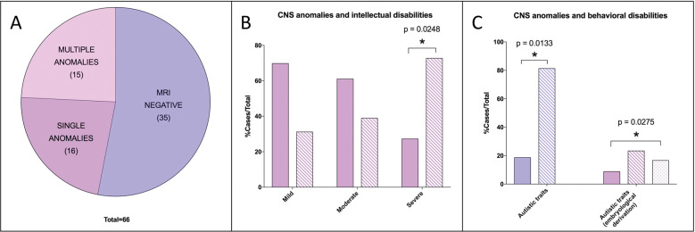
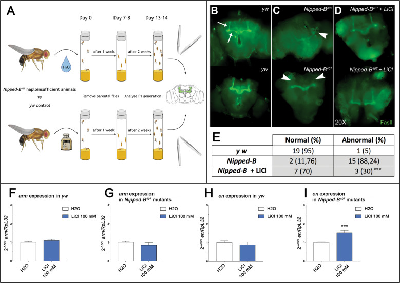
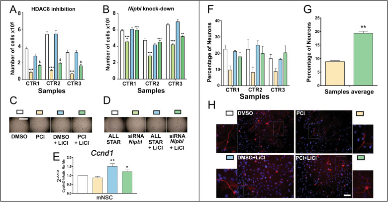
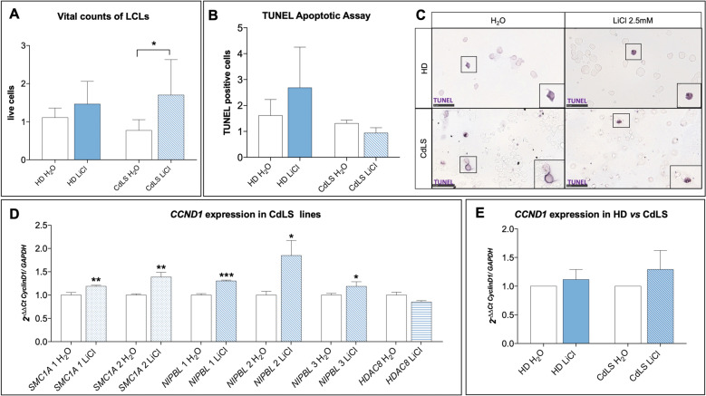
Cornelia de Lange Syndrome (CdLS) is a rare developmental disorder affecting a multitude of organs including the central nervous system, inducing a variable neurodevelopmental delay. CdLS malformations derive from the deregulation of developmental pathways, inclusive of the canonical WNT pathway. We have evaluated MRI anomalies and behavioral and neurological clinical manifestations in CdLS patients. Importantly, we observed in our cohort a significant association between behavioral disturbance and structural abnormalities in brain structures of hindbrain embryonic origin. Considering the cumulative evidence on the cohesin-WNT-hindbrain shaping cascade, we have explored possible ameliorative effects of chemical activation of the canonical WNT pathway with lithium chloride in different models: (I) Drosophila melanogaster CdLS model showing a significant rescue of mushroom bodies morphology in the adult flies; (II) mouse neural stem cells restoring physiological levels in proliferation rate and differentiation capabilities toward the neuronal lineage; (III) lymphoblastoid cell lines from CdLS patients and healthy donors restoring cellular proliferation rate and inducing the expression of CyclinD1. This work supports a role for WNT-pathway regulation of CdLS brain and behavioral abnormalities and a consistent phenotype rescue by lithium in experimental models.
Conflict of interest statement
The study is in accordance with the 1964 Helsinki declaration and its later amendments or comparable ethical standards. Written informed consent of patients or caregivers were collected for biological samples studies.
Figures
Similar articles
-
Integrating molecular and structural findings: Wnt as a possible actor in shaping cognitive impairment in Cornelia de Lange syndrome.Orphanet J Rare Dis. 2017 Nov 21;12(1):174. doi: 10.1186/s13023-017-0723-0. Orphanet J Rare Dis. 2017. PMID: 29162129 Free PMC article. Review.
-
Cornelia de Lange syndrome: Correlation of brain MRI findings with behavioral assessment.Am J Med Genet C Semin Med Genet. 2016 Jun;172(2):190-7. doi: 10.1002/ajmg.c.31503. Epub 2016 May 10. Am J Med Genet C Semin Med Genet. 2016. PMID: 27164360 Free PMC article.
-
Cornelia de Lange syndrome, related disorders, and the Cohesin complex: Abstracts from the 8th biennial scientific and educational symposium 2018.Am J Med Genet A. 2019 Jun;179(6):1080-1090. doi: 10.1002/ajmg.a.61108. Epub 2019 Mar 15. Am J Med Genet A. 2019. PMID: 39070592 Free PMC article.
-
Impairment of Retinoic Acid Signaling in Cornelia de Lange Syndrome Fibroblasts.Birth Defects Res. 2017 Oct 2;109(16):1268-1276. doi: 10.1002/bdr2.1070. Epub 2017 Jul 28. Birth Defects Res. 2017. PMID: 28752682
-
Cornelia de Lange Syndrome as Paradigm of Chromatinopathies.Front Neurosci. 2021 Nov 5;15:774950. doi: 10.3389/fnins.2021.774950. eCollection 2021. Front Neurosci. 2021. PMID: 34803598 Free PMC article. Review.
Cited by
-
Evidence of SARS-CoV-2 in nasal brushings and olfactory mucosa biopsies of COVID-19 patients.PLoS One. 2022 Apr 12;17(4):e0266740. doi: 10.1371/journal.pone.0266740. eCollection 2022. PLoS One. 2022. PMID: 35413071 Free PMC article.
-
The Designer Drug αPHP Affected Cell Proliferation and Triggered Deathly Mechanisms in Murine Neural Stem/Progenitor Cells.Biology (Basel). 2023 Sep 11;12(9):1225. doi: 10.3390/biology12091225. Biology (Basel). 2023. PMID: 37759624 Free PMC article.
-
NSC Physiological Features in Spinal Muscular Atrophy: SMN Deficiency Effects on Neurogenesis.Int J Mol Sci. 2022 Dec 2;23(23):15209. doi: 10.3390/ijms232315209. Int J Mol Sci. 2022. PMID: 36499528 Free PMC article. Review.
-
Cohesin Mutations Induce Chromatin Conformation Perturbation of the H19/IGF2 Imprinted Region and Gene Expression Dysregulation in Cornelia de Lange Syndrome Cell Lines.Biomolecules. 2021 Nov 2;11(11):1622. doi: 10.3390/biom11111622. Biomolecules. 2021. PMID: 34827619 Free PMC article.
-
Exploring the uncharted role of cell senescence in rare diseases.Orphanet J Rare Dis. 2025 Sep 1;20(1):465. doi: 10.1186/s13023-025-03778-1. Orphanet J Rare Dis. 2025. PMID: 40890826 Free PMC article. Review.
References
-
- Sinkey, R. G., Odibo, A. O. & Bradshaw, R. J. Cornelia de Lange syndrome. Obstetric Imaging: Fetal Diagnosis and Care, 2nd edn (University of Washington, Seattle, 2017). 10.1016/B978-0-323-44548-1.00127-3
Grants and funding
- 2015-0783/Fondazione Cariplo (Cariplo Foundation)
- 20661/Associazione Italiana per la Ricerca sul Cancro (Italian Association for Cancer Research)
- 20661/Associazione Italiana per la Ricerca sul Cancro (Italian Association for Cancer Research)
- 18-0399/AICR_/Worldwide Cancer Research/United Kingdom
- 18-0399/AICR_/Worldwide Cancer Research/United Kingdom
LinkOut - more resources
Full Text Sources
Other Literature Sources
Medical
Molecular Biology Databases
Research Materials
