Perineuronal nets are under the control of type-5 metabotropic glutamate receptors in the developing somatosensory cortex
- PMID: 33597513
- PMCID: PMC7889908
- DOI: 10.1038/s41398-021-01210-3
Perineuronal nets are under the control of type-5 metabotropic glutamate receptors in the developing somatosensory cortex
Abstract
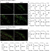
mGlu5 metabotropic glutamate receptors are highly functional in the early postnatal life, and regulate developmental plasticity of parvalbumin-positive (PV+) interneurons in the cerebral cortex. PV+ cells are enwrapped by perineuronal nets (PNNs) at the closure of critical windows of cortical plasticity. Changes in PNNs have been associated with neurodevelopmental disorders. We found that the number of Wisteria Fluoribunda Agglutinin (WFA)+ PNNs and the density of WFA+/PV+ cells were largely increased in the somatosensory cortex of mGlu5-/- mice at PND16. An increased WFA+ PNN density was also observed after pharmacological blockade of mGlu5 receptors in the first two postnatal weeks. The number of WFA+ PNNs in mGlu5-/- mice was close to a plateau at PND16, whereas continued to increase in wild-type mice, and there was no difference between the two genotypes at PND21 and PND60. mGlu5-/- mice at PND16 showed increases in the transcripts of genes involved in PNN formation and a reduced expression and activity of type-9 matrix metalloproteinase in the somatosensory cortex suggesting that mGlu5 receptors control both PNN formation and degradation. Finally, unilateral whisker stimulation from PND9 to PND16 enhanced WFA+ PNN density in the contralateral somatosensory cortex only in mGlu5+/+ mice, whereas whisker trimming from PND9 to PND16 reduced WFA+ PNN density exclusively in mGlu5-/- mice, suggesting that mGlu5 receptors shape the PNN response to sensory experience. These findings disclose a novel undescribed mechanism of PNN regulation, and lay the groundwork for the study of mGlu5 receptors and PNNs in neurodevelopmental disorders.
Conflict of interest statement
The authors declare that they have no conflict of interest.
Figures


Similar articles
-
A type-5 metabotropic glutamate receptor-perineuronal net axis shapes the function of cortical GABAergic interneurons in chronic pain.J Anesth Analg Crit Care. 2025 Feb 21;5(1):10. doi: 10.1186/s44158-025-00228-z. J Anesth Analg Crit Care. 2025. PMID: 39985105 Free PMC article. Review.
-
Sensory experience-dependent formation of perineuronal nets and expression of Cat-315 immunoreactive components in the mouse somatosensory cortex.Neuroscience. 2017 Jul 4;355:161-174. doi: 10.1016/j.neuroscience.2017.04.041. Epub 2017 May 8. Neuroscience. 2017. PMID: 28495333
-
A Progressive Build-up of Perineuronal Nets in the Somatosensory Cortex Is Associated with the Development of Chronic Pain in Mice.J Neurosci. 2022 Apr 6;42(14):3037-3048. doi: 10.1523/JNEUROSCI.1714-21.2022. Epub 2022 Feb 22. J Neurosci. 2022. PMID: 35193928 Free PMC article.
-
Experience-dependent development of perineuronal nets and chondroitin sulfate proteoglycan receptors in mouse visual cortex.Matrix Biol. 2013 Aug 8;32(6):352-63. doi: 10.1016/j.matbio.2013.04.001. Epub 2013 Apr 15. Matrix Biol. 2013. PMID: 23597636
-
The Perineuronal 'Safety' Net? Perineuronal Net Abnormalities in Neurological Disorders.Front Mol Neurosci. 2018 Aug 3;11:270. doi: 10.3389/fnmol.2018.00270. eCollection 2018. Front Mol Neurosci. 2018. PMID: 30123106 Free PMC article. Review.
Cited by
-
Neural Correlates of Auditory Hypersensitivity in Fragile X Syndrome.Front Psychiatry. 2021 Oct 7;12:720752. doi: 10.3389/fpsyt.2021.720752. eCollection 2021. Front Psychiatry. 2021. PMID: 34690832 Free PMC article. Review.
-
Developmental up-regulation of NMDA receptors in the prefrontal cortex and hippocampus of mGlu5 receptor knock-out mice.Mol Brain. 2021 May 7;14(1):77. doi: 10.1186/s13041-021-00784-9. Mol Brain. 2021. PMID: 33962661 Free PMC article.
-
Molecular and Cellular Insights: A Focus on Glycans and the HNK1 Epitope in Autism Spectrum Disorder.Int J Mol Sci. 2023 Oct 13;24(20):15139. doi: 10.3390/ijms242015139. Int J Mol Sci. 2023. PMID: 37894820 Free PMC article.
-
Parvalbumin interneuron-derived tissue-type plasminogen activator shapes perineuronal net structure.BMC Biol. 2022 Oct 5;20(1):218. doi: 10.1186/s12915-022-01419-8. BMC Biol. 2022. PMID: 36199089 Free PMC article.
-
A type-5 metabotropic glutamate receptor-perineuronal net axis shapes the function of cortical GABAergic interneurons in chronic pain.J Anesth Analg Crit Care. 2025 Feb 21;5(1):10. doi: 10.1186/s44158-025-00228-z. J Anesth Analg Crit Care. 2025. PMID: 39985105 Free PMC article. Review.
References
Publication types
MeSH terms
Substances
LinkOut - more resources
Full Text Sources
Other Literature Sources

