. 2021 Feb 17;4(1):226.
doi: 10.1038/s42003-021-01750-w.

The structural basis of function and regulation of neuronal cotransporters NKCC1 and KCC2

Affiliations

The structural basis of function and regulation of neuronal cotransporters NKCC1 and KCC2

Sensen Zhang et al. Commun Biol. .

Abstract

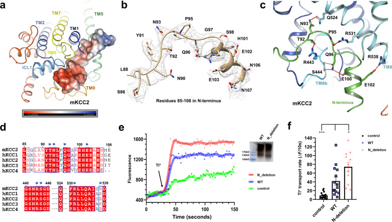
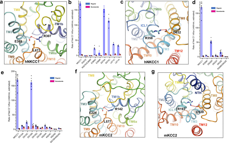
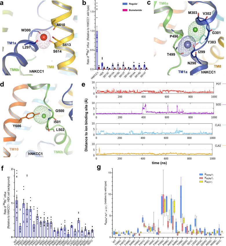
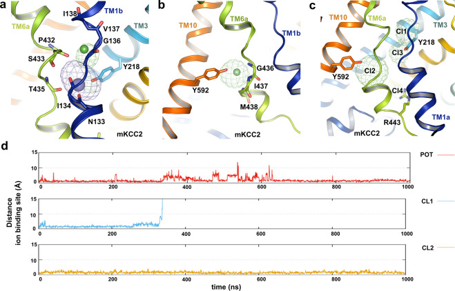
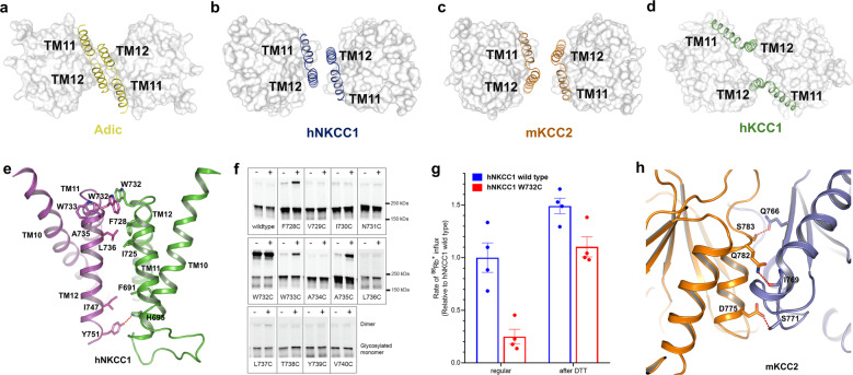
NKCC and KCC transporters mediate coupled transport of Na++K++Cl- and K++Cl- across the plasma membrane, thus regulating cell Cl- concentration and cell volume and playing critical roles in transepithelial salt and water transport and in neuronal excitability. The function of these transporters has been intensively studied, but a mechanistic understanding has awaited structural studies of the transporters. Here, we present the cryo-electron microscopy (cryo-EM) structures of the two neuronal cation-chloride cotransporters human NKCC1 (SLC12A2) and mouse KCC2 (SLC12A5), along with computational analysis and functional characterization. These structures highlight essential residues in ion transport and allow us to propose mechanisms by which phosphorylation regulates transport activity.


Conflict of interest statement

The authors declare no competing interests.

Figures

Fig. 1
Fig. 1. Structures of hNKCC1 and mKCC2.
a Cryo-EM map of the high-resolution hNKCC1 from a side view with each subunit color-coded (blue and green). b Ribbon representation of the high-resolution hNKCC1 from a side view with each subunit color-coded (blue and green). c The TM-domain of the DrNKCC1 (red), previous reported hNKCC1(K289N_G351R) (brown), and the current hNKCC1 (blue) NKCC1 single monomers are superimposed. Top view (from the extracellular side). d Cryo-EM map of the dimeric mKCC2 from a side view with each subunit color-coded (yellow and brown). e Ribbon representation of the dimeric mKCC2 from a side view with each subunit color-coded (yellow and brown). f Superimposition of single monomers of hKCC1 (green) and mKCC2 (yellow) in the TMD demonstrates good alignment (RMSD of 0.69 Å over 341 Cα atoms) except for the TM12 helix which is tilted 23° in hKCC1 compared to mKCC2.
Fig. 2
Fig. 2. Inhibitory N-terminus peptide in mKCC2.
a An N-terminus peptide fragment blocks the entrance to the translocation pore of mKCC2. The peptide is shown as the electrostatic surface potential from -5 to +5 kT/e. b Density map of the representative residues 85–108 in N terminus of mKCC2 showing the 10.0 σ contour. Representative atomic model (gold) with side chains was fitted into the cryo-EM density map (gray mesh). c Interactions between the N-terminus peptide and TM domains of mKCC2. T92, Q96, N93, P95, and E102 from the N-terminus peptide interact with R443 and S444 from TM6b, and interact with Q524, R531, and R538 from TM8 in mKCC2. d Sequence alignment KCCs in the regions of interaction between the N terminus, and of TMs 6 and 8. Numbering is that of mKCC2b. Interacting residues are highlighted by blue labels. e Fluorescence signal measurement of the cells transfected by control (mock virus), WT, and N-deletion (residue 85–120 deleted) mKCC2 virus. Transport activity was initiated by the addition of Tl+ (arrow). The inset western-blot shows an expression of WT and N-(85–120) deleted mKCC2. f Relative Tl+ transport rate in control (n = 17), WT (n = 14), and N-deletion (n = 12) mKCC2. Unpaired Student’s t-test was used to evaluate the significance between WT and N-deletion mKCC2 (*P < 0.05), and between WT and control (***P < 0.001); mean ± SEM.
Fig. 3
Fig. 3. Potential gating residues in hNKCC1 and mKCC2.
a The amino group of R307 of TM1b interacts with the carbonyl oxygen of E389 from TM3 to constrict the extracellular gate of hNKCC1. b hNKCC1 transport activity is greatly decreased by mutation of the extracellular gating amino acids E389, R307, and L671; mean ± SEM. c At the intracellular end of the hNKCC1 translocation pore, R358 of ICL1 interacts with D632 of the neighboring TM8. d hNKCC1 transport activity between WT and mutants in the intracellular essential amino acids (R358 and D632); mean ± SEM. e hNKCC1 mutants of D510 and K624 at the intracellular end of the pore have greatly reduced transport activity—these residues are implicated in MD simulations (see also Supplementary Fig. 10a, b); mean ± SEM. f Homologous to hNKCC1 (a), the extracellular gate of mKCC2 is restricted by hydrogen bonds between R142 of TM1b and E224 of TM3. g Potential intracellular-gating residues of mKCC2: homologous to hNKCC1 (c), R193 of ICL1 in mKCC2 is close to D539 of TM8.
Fig. 4
Fig. 4. Ion-binding probability densities and mutagenesis assay of hNKCC1.
a The Na+ binding site of hNKCC1, based on molecular dynamic simulations. Na+ is stabilized by interactions with S613, S614, and A610 in TM8, and L297 and W300 in TM1. b hNKCC1 transport activity is greatly decreased by mutations of S613 and S614 in the proposed Na+-binding site; mean ± SEM. c K+ and Cl (Cl-1) binding sites of hNKCC1 based on molecular dynamic simulations. The K+ ion is stabilized by interaction with Y383 from TM3, N298, and I299 in TM1, P496, and T499 in TM6 and by the Cl ion. The Cl ion is stabilized by interaction with G301, V302, and M303 from TM1 and by the K+ ion. d A second Cl binding site in hNKCC1 (Cl-2), based on molecular dynamic simulations. This Cl ion is stabilized by interactions with G500, I501, and L502 in TM6a, and Y686 in TM10. e Time evolution of the distance of the four ions (Na+, K+, Cl-1, and Cl-2) from its proposed binding sites based on molecular dynamics simulation. f hNKCC1 86Rb+ transport activity of cysteine mutants in a scan of TM6; mean ± SEM. g The relative KM (for Na+, Rb+, and Cl) for activation of transport of each of the cysteine mutants in TM6 as a ratio to the wild-type hNKCC1. The absolute KMs of wild-type hNKCC1 were 5.6, 3.3, and 18.3 mM for Na+, Rb+, and Cl, respectively. “Whiskers” indicate the maximum range of the values.
Fig. 5
Fig. 5. Ion-binding probability densities of mKCC2.
a K+ and Cl (Cl-1) binding sites of mKCC2 based on molecular dynamic simulations. Similar to hNKCC1, K+ is stabilized by the interaction with Y218 in TM3, N133 and I134 in TM1a, T435 and P432 in TM6, and the Cl ion. The Cl binding site is stabilized by interaction with G136, V137, and I138 in TM1 and by the K+ ion. b A second Cl binding site (Cl-2) of mKCC2. Similar to (Cl-2) in hNKCC1, this Cl is stabilized by the interaction with G436, I437, and M438 in TM6, and Y592 in TM10. c Four Cl ion probability densities in mKCC2 based on molecular dynamic simulations. d Time evolution of the distance of the three ions (K+, Cl-1, and Cl-2) from its proposed binding sites based on molecular dynamics simulation. The Cl ion escapes from the Cl-1 site to the neighboring Cl-3 site after 300 ns.
Fig. 6
Fig. 6. Dimeric interface of hNKCC1 and mKCC2.
ad Schematic representation of the TMD dimer architecture between different transporters (Adic (yellow), hNKCC1 (blue), mKCC2 (brown), and hKCC1 (green)) with TM11-12 shown as ribbons and other TMD regions shown as surface representation. e The NKCC1 homodimer is stabilized by hydrogen bond interaction between Y751 in TM12 of one monomer with H695 in TM10 of the other; in addition, there are stabilizing hydrophobic interactions between TM11-12 hairpins (labeled residues). f Homodimeric oxidative crosslinking of NKCC1 with single cysteine substitutions in TM 11-12. Cells were treated with (+) or without (−) cupric phenanthroline before lysis. g The W732C mutant exhibits about 25% of the Rb+ influx activity of wild-type NKCC1. Transport activity is largely restored in W732C after a 30 min pretreatment with 40 mM DTT to break the disulfide linkage between hNKCC1 monomers; mean ± SEM. h Cytoplasmic-domain dimeric interactions in mKCC2. D775, Q782, and S783 from one subunit (brown) form hydrogen bonds with Q766, I769, and S771 from the neighboring subunit (blue) to stabilize the dimer.
Fig. 7
Fig. 7. Hypotheses of regulatory activation in CCCs.
a The basic model of cation-chloride cotransporter activation. It is proposed that the inactive state is characterized by an inward-open TM conformation with CTDs in a tightly organized domain-swap configuration. On activation, the CTDs are released to a freely tethered configuration with accompanying change in the dimer architecture. b Our hypothesis is that NKCC activation occurs when the phosphorylated N terminus interacts with the CTD and disrupts the dimer. c The KCC inactive state exhibits an N terminus inhibitory peptide blocking the transport pore. Activation occurs when the peptide instead binds to the non-phosphorylated C terminus.

Similar articles

Cited by

References

    1. Russell JM. Sodium-potassium-chloride cotransport. Physiol. Rev. 2000;80:211–276. doi: 10.1152/physrev.2000.80.1.211. - DOI - PubMed
    1. Haas M, Forbush B. The Na-K-Cl cotransporter of secretory epithelia. Annu. Rev. Physiol. 2000;62:515–51. doi: 10.1146/annurev.physiol.62.1.515. - DOI - PubMed
    1. Kahle KT, et al. K-Cl cotransporters, cell volume homeostasis, and neurological disease. Trends Mol. Med. 2015;21:513–523. doi: 10.1016/j.molmed.2015.05.008. - DOI - PMC - PubMed
    1. Gamba G. Molecular physiology and pathophysiology of electroneutral cation-chloride cotransporters. Physiol. Rev. 2005;85:423–493. doi: 10.1152/physrev.00011.2004. - DOI - PubMed
    1. Payne JA. Molecular operation of the cation chloride cotransporters: ion binding and inhibitor interaction. Co. Transp. Syst. 2012;70:215–237. doi: 10.1016/B978-0-12-394316-3.00006-5. - DOI - PubMed

Publication types

MeSH terms

LinkOut - more resources