. 2021 Mar;591(7849):327-331.
doi: 10.1038/s41586-021-03240-9. Epub 2021 Feb 17.

Glutamate transporters have a chloride channel with two hydrophobic gates

Affiliations

Glutamate transporters have a chloride channel with two hydrophobic gates

Ichia Chen et al. Nature. 2021 Mar.

Abstract

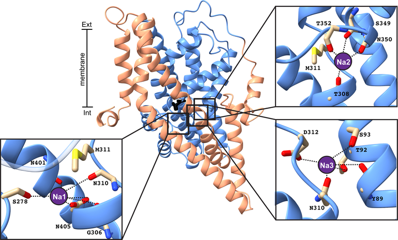
Glutamate is the most abundant excitatory neurotransmitter in the central nervous system, and its precise control is vital to maintain normal brain function and to prevent excitotoxicity1. The removal of extracellular glutamate is achieved by plasma-membrane-bound transporters, which couple glutamate transport to sodium, potassium and pH gradients using an elevator mechanism2-5. Glutamate transporters also conduct chloride ions by means of a channel-like process that is thermodynamically uncoupled from transport6-8. However, the molecular mechanisms that enable these dual-function transporters to carry out two seemingly contradictory roles are unknown. Here we report the cryo-electron microscopy structure of a glutamate transporter homologue in an open-channel state, which reveals an aqueous cavity that is formed during the glutamate transport cycle. The functional properties of this cavity, combined with molecular dynamics simulations, reveal it to be an aqueous-accessible chloride permeation pathway that is gated by two hydrophobic regions and is conserved across mammalian and archaeal glutamate transporters. Our findings provide insight into the mechanism by which glutamate transporters support their dual function, and add information that will assist in mapping the complete transport cycle shared by the solute carrier 1A transporter family.


Conflict of interest statement

Competing interest declaration

The authors declare no competing interests.

Figures

Extended Data Fig. 1 |
Extended Data Fig. 1 |. Cross-linking experiments on purified GltPh double cysteine transporters.
a, SDS-PAGE gel shift assay shows the extent of cross-linking in detergent-solubilized cysteine-less GltPh (CLGltPh) and the double cysteine GltPh transporters under untreated conditions, upon mPEG5K-maleimide treatment and following incubation with HgCl2 with arrows indicating the positions of differentially cross-linked protomers and mPEG5K-bound proteins. This is representative data from one experiment that was replicated at least two times from two separate protein purifications. For gel source data, see Supplementary Figure 1. b, Crystal structure of GltPh-XL1 (purple) and GltPh-XL3 (pink) superimposed on the OFS (PDB: 2NWX; left, grey) and the IFS (PDB: 3KBC; right, grey), respectively. c, SDS-PAGE analysis of purified GltPh-XL3 in nanodiscs. d, Cryo-EM structure of GltPh-XL2 (green) superimposed on the iOFS (PDB:3V8G, chain C; grey).
Extended Data Fig. 2 |
Extended Data Fig. 2 |. Cryo-EM data processing protocol and refinement.
a, Data processing flow chart for GltPh reconstituted into nanodiscs in the presence of NaCl and aspartate. b, Fourier shell correlation (FSC) curves indicating the resolution at the 0.143 threshold of final masked (blue) and unmasked (red) maps for GltPh trimer iOFS (left), GltPh protomer ClCS (middle) and GltPh protomer iOFS (right). c, Final maps after Relion post-processing, colored according to local resolution estimation using Relion for GltPh trimer iOFS (left, 3.9Å resolution, contour level 7.2σ), GltPh protomer ClCS (middle, 4.0Å resolution, contour level 12.0σ) and GltPh protomer iOFS (right, 3.7Å resolution, contour level 9.5σ). Contour levels calculated using Chimera.
Extended Data Fig. 3 |
Extended Data Fig. 3 |. The conformational space of a GltPh protomer.
a, The front and b, top views of the cryo-EM map and atomic model of GltPh-XL2 in the iOFS (contour level 9.5σ) and ClCS (contour level 12.0σ). Density attributed to the scaffold domain, transport domain and HP2 are shown in salmon, blue and red, respectively. c, Conformational changes undertaken by a GltPh protomer during the substrate transport cycle viewed from the side and top. HP2 is colored for easier visualization of rotational changes observed in the transport domain. d, e, Close-up views of the L152C-G351C cross-link fitted in the iOFS (contour level 12.9σ) and ClCS (contour level 10.3σ) cryo-EM maps. Contour level calculated using Chimera.
Extended Data Fig. 4 |
Extended Data Fig. 4 |. Na+ coordination sites in the ClCS.
A close-up of the three Na+ coordination sites on the ClCS protomer are shown with residues interacting with Na+ ion (purple circle, modelled) shown in sticks. The scaffold and the transport domains are shown in salmon and blue, respectively, with the substrate in black sticks.
Extended Data Fig. 5 |
Extended Data Fig. 5 |. Nanodisc deformation supports transport domain movement by the ClCS and putative lipid binding sites.
a, Percentage of GltPh-XL2 trimers containing all three protomers in iOFS, in ClCS, or a mixture of both. Out of 220,938 trimers, 79,809 contained one or more protomers in the ClCS. Particle count within symmetry expanded data showed 63,470 trimers contained one ClCS, 14,394 trimers contained two ClCS and 1,945 trimers contained all protomers in the ClCS. b, Density map of GltPh-XL2 trimer (unfocused refinement) containing one ClCS protomer and embedded in nanodiscs (viewed from the membrane plane). The two iOFS protomers are shown in orange and the ClCS in blue. The nanodisc is shown in yellow. c, Putative lipid binding sites in the GltPh-XL2 trimer, where the transport domain is in blue and the scaffold domain in salmon. Identical lipid densities (green) were observed between protomers (contour level 7.2σ). TMs located within 5 Å of the putative lipid densities are labelled. Contour level calculated using Chimera.
Extended Data Fig. 6 |
Extended Data Fig. 6 |. Water conduction through GltPh-ClCS and setup of umbrella sampling simulations and convergence to capture Cl movement through GltPh-ClCS.
a, The GltPh-ClCS structure was embedded into a lipid bilayer containing PE, PG, and PC lipids (mimicking experimental conditions). After an initial equilibration of 100 ns, the entire system was subjected to an external electric field of 800mV which resulted in a continuous water pathway through the interface of scaffold and transport domain. The GltPh-ClCS protomer is shown in cartoon, with the transport domain in blue and the scaffold domain in salmon. XL-2 residues L152/G351 are shown in red and blue spheres, respectively b, Residues lining the Cl pathway have a higher solvent accessible surface area (SASA) in the GltPh-ClCS than in the OFS (calculated using the crystal structure of OFS PDB: 2NWX). c, Overlap between the corresponding windows used in US simulations. d, No significant changes between the free-energy profile obtained at 10ns (red), 15ns (blue), and 20 ns (green) were observed, highlighting convergence of US simulations.
Extended Data Fig. 7 |
Extended Data Fig. 7 |. The EAAT1 open-channel conformation conducts Cl.
a, L-[3H]glutamate uptake into oocytes expressing cysteine-less EAAT1 and double cysteine transporter mutants in control conditions (grey), and following pre-incubation with DTT (cyan) or CuPh (orange). Number of cells (n) used for each condition is indicated in each graph and all measurements presented were taken across at least two batches of oocytes. b-e, L-glutamate elicited current-voltage relationships for cysteine-less E1 (b), E1-XL1 (c), E1-XL2 (d) and E1-XL3 (e) monitored in the same conditions as (a). To confirm cross-links E1-XL1, E1-XL2 and E1-XL3 were occurring within an individual protomer rather than between protomers of the trimeric complex, oocytes expressing single cysteine residues that make up E1-XL1 (K300C and W473C), E1-XL2 (L244C and G439C), and E1-XL3 (K300C and A470C) either alone or co-injected into an individual oocyte were also examined using the same approaches (f-g). Data represent standard error of the mean (mean ± SEM). h, hEAAT1 (PBD: 5LLU) highlighting residues forming the extracellular and intracellular hydrophobic gates. The scaffold domain is shown in grey and the transport domain in gold. The Cα atoms of the two introduced cysteine residues are shown as spheres (L244 in red and G439 in blue). i, Membrane reversal potentials (Erev) measured in oocytes expressing wild-type (n = 6) and mutant (n = 5) EAAT1 transporters. Each white circle represents a response from a single cell. Black bar represents mean ± SEM. Significance was determined using One-Way ANOVA with Bonferroni post-hoc test for multiple comparisons performed by GraphPad Prism 8, and exact P values are provided. j, Schematic representation of the substrate transport cycle. A single protomer is shown with the scaffold domain in salmon, transport domain in blue and substrate in black.
Fig. 1 |
Fig. 1 |. GltPh utilizes an elevator mechanism that can be probed by cross-links between the transport and scaffold domains.
a, GltPh protomer viewed parallel to the membrane in the OFS (PDB:2NWX), iOFS (PDB:3V8G) and IFS (PDB:3KBC) with the scaffold domain (salmon), the transport domain (blue) and bound aspartate (black). The three cysteine pairs, XL1, XL2 and XL3, are in purple, green and pink, respectively. b, Cα-Cα distances of cysteine pairs in the known crystal structures of GltPh.
Fig. 2 |
Fig. 2 |. GltPh can be trapped in an open-channel conformation.
a, Surface representation of GltPh-XL1 in the OFS, GltPh-XL2 in the iOFS and ClCS, and GltPh-XL3 in the IFS, viewed from the membrane plane. The color scheme is the same as in Fig. 1. The Cα positions of L152C and G351C are shown as spheres in red and blue, respectively. The dashed line through L152C highlights its unchanging position throughout the transport cycle as G351C moves down. b, Cross-sections of (a) through R276. Residues lining the domain interface are labelled. Close-up of the “constriction zone” viewed from the extracellular space in the iOFS (contour level 12.9σ) (c) and the ClCS (contour level 10.3σ) (d) fitted in their respective cryo-EM maps. e, Close-up of the narrowest point of the cavity in the ClCS (contour level 10.3σ). Noise removed for clarity; contour levels calculated using Chimera.
Fig. 3 |
Fig. 3 |. Energetic landscape for the movement of Cl through GltPh-ClCS.
a, The ClCS structure was embedded into a lipid bilayer and simulated for 300 ns with and without an external electric field during which a continuous water pathway (red surface) formed at the domain interface. b, The inter-domain water molecules were used to place individual Cl ions (purple spheres) to seed the windows for US simulations that resulted in the underlying free-energy profile for Cl conduction. c, Occupancy of Cl captured during 1.2-μs US simulation is shown in light red isosurface. Residues which form the Cl pathway (polar (green), hydrophobic (cyan), basic (blue), acidic (red)) are labeled. The ClCS protomer is shown in cartoon, scaffold domain (salmon) and transport domain (blue). d, The free energy profile for Cl conduction through ClCS along the pore axis, obtained from the US simulations, shows favorable interactions (minima) between the Cl ion and R276/R52/K55, and energy barriers formed by hydrophobic residues as indicated. Overall, the largest barrier against the movement of Cl observed is 1.9 kcal/mol. e, Residues interacting with the permeating Cl ion.

Similar articles

Cited by

References

    1. Vandenberg RJ, Ryan RM, Mechanisms of glutamate transport. Physiol Rev 93, 1621–1657 (2013). - PubMed
    1. Reyes N, Ginter C, Boudker O, Transport mechanism of a bacterial homologue of glutamate transporters. Nature 462, 880–885 (2009). - PMC - PubMed
    1. Ryan RM, Vandenberg RJ, Elevating the alternating-access model. Nat Struct Mol Biol 23, 187–189 (2016). - PubMed
    1. Zerangue N, Kavanaugh MP, Flux coupling in a neuronal glutamate transporter. Nature 383, 634–637 (1996). - PubMed
    1. Wadiche JI, Arriza JL, Amara SG, Kavanaugh MP, Kinetics of a human glutamate transporter. Neuron 14, 1019–1027 (1995). - PubMed

Publication types

MeSH terms

LinkOut - more resources