. 2021 Feb 1:11:600227.
doi: 10.3389/fgene.2020.600227. eCollection 2020.

CDK6 Is a Potential Prognostic Biomarker in Acute Myeloid Leukemia

Affiliations

CDK6 Is a Potential Prognostic Biomarker in Acute Myeloid Leukemia

Wei Liu et al. Front Genet. .

Abstract

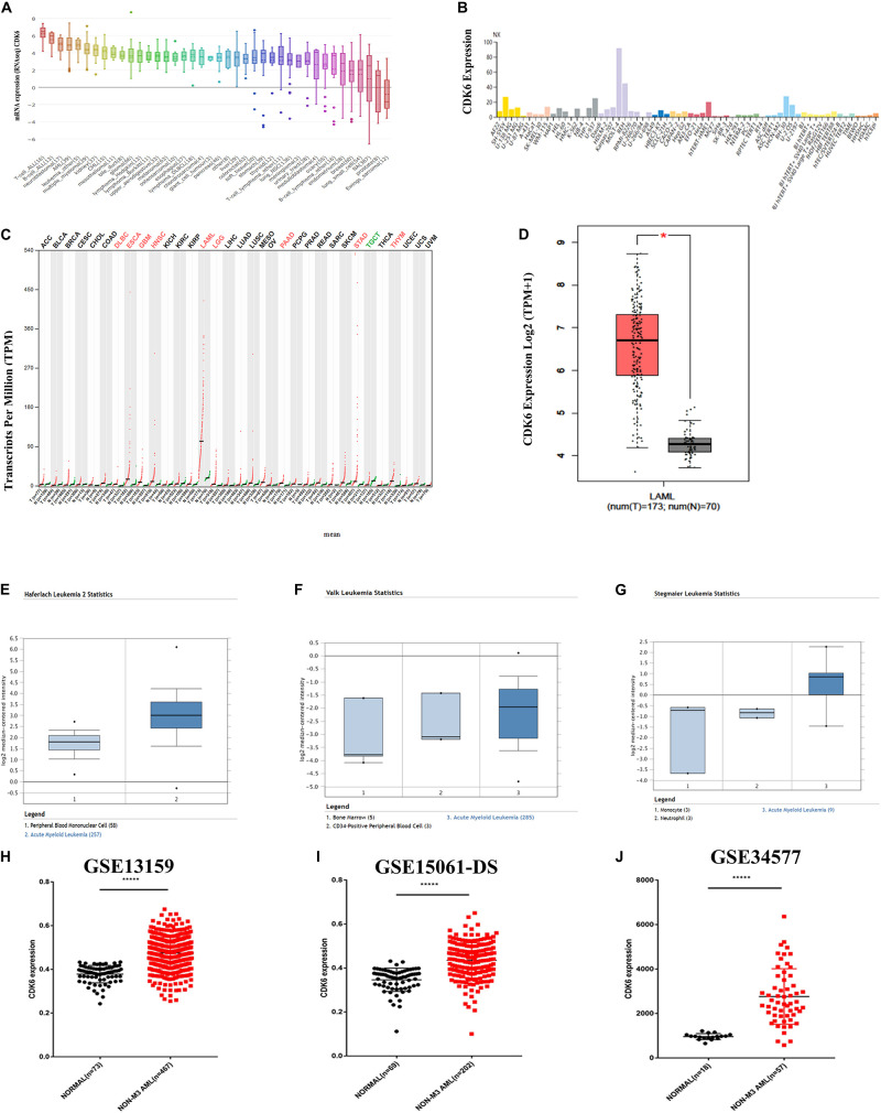
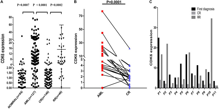
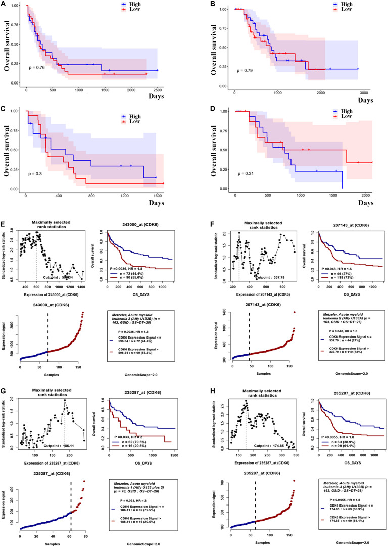
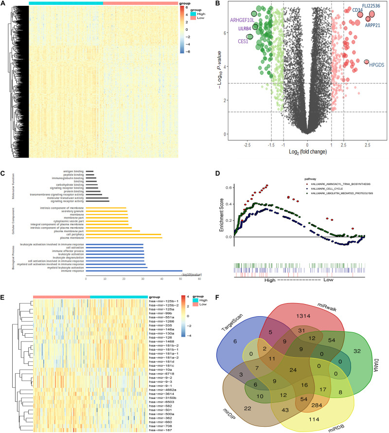
Acute myeloid leukemia (AML) is a threatening hematological malignant disease in which new successful approaches in therapy are needed. Cyclin-dependent kinase 6 (CDK6), a regulatory enzyme of the cell cycle that plays an important role in leukemogenesis and the maintenance of leukemia stem cells (LSC), has the potential to predict the prognosis of AML. By analyzing public databases, we observed that the messenger RNA (mRNA) levels of CDK6 were significantly overexpressed in AML cell lines and non-acute promyelocytic leukemia (non-APL) AML patients when compared to healthy donors. Furthermore, CDK6 expression was significantly reduced in AML patients who achieved complete remission (CR) compared to that at the time of diagnosis in our validated cohort. The expression of CDK6 was tightly correlated with peripheral blood blasts, French-American-British (FAB) subtypes, CCAAT-enhancer-binding protein α (CEBPA) mutation, and chromosomal abnormalities of t(8;21). However, the clinical significance and effects of CDK6 expression on the prognosis of non-APL AML patients remain uncertain. We found that CDK6 expression was inversely correlated with overall survival (OS) among non-APL AML patients using the Kaplan-Meier analysis. CDK6 was also found to be positively associated with genes identified to contribute to the development of leukemia, including CCND2, DNMT3B, SOX4, and IKZF2, as well as being negatively associated with anticancer microRNAs, including miR-187, miR-9, miR-582, miR708, and miR-362. In summary, our study revealed that CDK6 might be a potential diagnostic and prognostic biomarker in non-APL AML patients.

Keywords: AML; CDK6; expression; prognosis; target therapy.


Conflict of interest statement

The authors declare that the research was conducted in the absence of any commercial or financial relationships that could be construed as a potential conflict of interest.

Figures

FIGURE 1
FIGURE 1
CDK6 overexpression in non-APL AML in the public database. (A) The expression of CDK6 in 40 types of human cancer cell line in the Cancer Cell Line Encyclopedia (CCLE) database. (B) The expression of CDK6 in human cancer cell lines in Human Protein Atlas (HPA) database. (C) The expression of CDK6 in 33 types compared with normal subjects in Gene Expression Profiling Interactive Analysis (GEPIA) database. (D) The expression of CDK6 in AML patients (n = 173) from TCGA and normal (n = 70) from GTX database by using (GEPIA) (p < 0.01). (E–G) In Haferlach leukemia2, Valk Leukemia, and Stegmaier leukemia statistics, CDK6 was overexpressed in AML patients than normal with a fold change = 2.388, p = 2.88E-30, fold change = 1.786, p = 0.025 and fold change = 3.179, p = 0.011, respectively. (H) The expression levels of CDK6 mRNA in non-APL AML patients(n = 467) compared with the normal samples (n = 73) (mean ± SEM level: 0.4748 ± 0.003325 vs. 0.3783 ± 0.004646, p < 0.0001) in GSE13159. (I) The expression level of CDK6 mRNA in non-APL AML patients (n = 202) compared with the normal samples (n = 69) (mean ± SEM level: 613.6 ± 30.5 vs. 253.2 ± 12.21, n = 69, p < 0.0001) in GSE15061-DS. (J) The expression level of CDK6 mRNA in non-APL AML patients (n = 57) compared with normal (n = 18) (mean ± SEM level: 961.7 ± 31.09 vs. 961.7 ± 31.09, p < 0.0001) in GSE34577.
FIGURE 2
FIGURE 2
Relative levels of CDK6 expression in the validated non-APL AML patients. (A) The CDK6 expression in healthy donors (n = 54), de novo non-APL patients (n = 127), AML-CR patients (n = 146), and AML-RR (n = 48) patients from Xiangya Hospital and The First Affiliated Hospital of Jinan University Hospital. The distributions of CDK6 expression were presented with scatter plots. The expression level of CDK6 mRNA in de novo non-APL AML patients (n = 127) compared with the normal samples (n = 54) (mean ± SEM level: 13.42 ± 1.46 vs. 1.75 ± 0.5703, p = 0.007). The expression level of CDK6 mRNA in de novo non-APL AML patients (n = 127) compared with AML-CR patients (n = 146) (mean ± SEM level: 13.42 ± 1.46 vs. 2.257 ± 0.2894, p < 0.0001). The expression level of CDK6 mRNA in AML-RR patients (n = 48) compared with AML-CR patients (n = 146) (mean ± SEM level: 15.11 ± 6.256 vs. 2.257 ± 0.2894, p = 0.0002). (B) The CDK6 expression at the time of diagnosis higher than at the time of evaluation for response following standard induction (3 + 7) chemotherapy in patients with AML who achieved CR (p = 0.0001) (n = paired groups of 29). (C) P1–P11 represented 11 paired AML non-APL patients with available follow-up data in first diagnosed time, complete remission (CR) and refractory remission (RR) time. (First diagnosed time vs. CR time: p = 0.002; RR vs. CR time: p = 0.0086, respectively) (p-Values were adjusted by the Holm–Sidak method. HD, healthy donor).
FIGURE 3
FIGURE 3
The effect of CDK6 expression on overall survival in non-APL AML patients. (A) Kaplan–Meier analysis of OS in chemotherapy only group in non-APL AML patients [CDK6high (n = 36) vs. CDK6low (n = 36): p = 0.76] (cut point: median CDK6 expression level). (B) Kaplan–Meier analysis of OS in auto-/allo-HSCT group in non-APL AML patients [CDK6high (n = 31) vs. CDK6low (n = 32): p = 0.79] (cut point: median CDK6 expression level). (C) Kaplan–Meier analysis of OS in chemotherapy only group in cytogenetically normal AML patients [CDK6high (n = 17) vs. CDK6low (n = 18): p = 0.3] (cut point: median CDK6 expression level). (D) Kaplan–Meier analysis of OS in auto-/allo-HSCT group in cytogenetically normal AML patients [CDK6high (n = 17) vs. CDK6low (n = 18): p = 0.3] (cut point: median CDK6 expression level). (E) The survival analysis of probe 243000_at of 162 cytogenetically normal AML (CN-AML) patients using the online web tool GenomicScape (cut point: 596.34, p = 0.0036, HR = 1.8); (F) the survival analysis of probe 207143_at of 162 cytogenetically normal AML (CN-AML) patient (cut point: 337.79, p = 0.048, HR = 1.6). (G) The survival analysis of probe 235287_at of 78 cytogenetically normal AML (CN-AML) patients using the online web tool Genomicscape (cut off: 596.34, p = 0.033, HR = 2). (H) The survival analysis of probe 235287_at of 162 cytogenetically normal AML (CN-AML) patients using the online web tool Genomicscape (cut point: 596.34, p = 0.0055, HR = 1.8).
FIGURE 4
FIGURE 4
Molecular signatures associated with CDK6 in non-APL AML. (A) Heatmap of differentially expressed genes (DEGs) between CDK6high and CDK6low non-APL AML patients. (B) Volcano plot of DEGs between CDK6high and CDK6low non-APL AML patients. (C) Biological process, molecular functions, and cellular component analysis of DEGs using the online website of Search Tool for the Retrieval of Interacting Genes/Proteins. (D) KEGG pathway analysis between CDK6high and CDK6low group by using the online tool Gene Set Enrichment Analysis (GSEA); the red dot represents aminoacyl tRNA biosynthesis pathway (NES = 1.78, p = 0.003), the blue dot represents the cell cycle pathway (NES = 1.35, p = 0.021), the green dot represents ubiquitin-mediated proteolysis pathway (NES = 1.64, p < 0.0001). (E) Heatmap of DEGs miRNAs CDK6high and CDK6low AML patients. (F) Venn results of microRNAs that could target CDK6 precited by online tools miRDB, miRwalk, TargetScan, mirDIP, and DIANA.

References

    1. Abdel-Wahab O., Levine R. L. (2013). Mutations in epigenetic modifiers in the pathogenesis and therapy of acute myeloid leukemia. Blood 121 3563–3572. 10.1182/blood-2013-01-451781 - DOI - PMC - PubMed
    1. Barrett T., Wilhite S. E., Ledoux P., Evangelista C., Kim I. F., Tomashevsky M., et al. (2013). NCBI GEO: archive for functional genomics data sets–update. Nucleic Acids Res. 41 D991–D995. 10.1093/nar/gks1193 - DOI - PMC - PubMed
    1. Chan F. K., Zhang J., Cheng L., Shapiro D. N., Winoto A. (1995). Identification of human and mouse p19, a novel CDK4 and CDK6 inhibitor with homology to p16ink4. Mol. Cell. Biol. 15 2682–2688. 10.1128/mcb.15.5.2682 - DOI - PMC - PubMed
    1. Chilosi M., Doglioni C., Yan Z., Lestani M., Menestrina F., Sorio C., et al. (1998). Differential expression of cyclin-dependent kinase 6 in cortical thymocytes and T-cell lymphoblastic lymphoma/leukemia. Am. J. Pathol. 152 209–217. - PMC - PubMed
    1. Deshpande A., Sicinski P., Hinds P. W. (2005). Cyclins and cdks in development and cancer: a perspective. Oncogene 24 2909–2915. 10.1038/sj.onc.1208618 - DOI - PubMed

LinkOut - more resources