. 2021 Feb 8;15(1):014108.
doi: 10.1063/5.0043312. eCollection 2021 Jan.

Creating wounds in cell monolayers using micro-jets

Affiliations

Creating wounds in cell monolayers using micro-jets

Cristian Soitu et al. Biomicrofluidics. .

Abstract

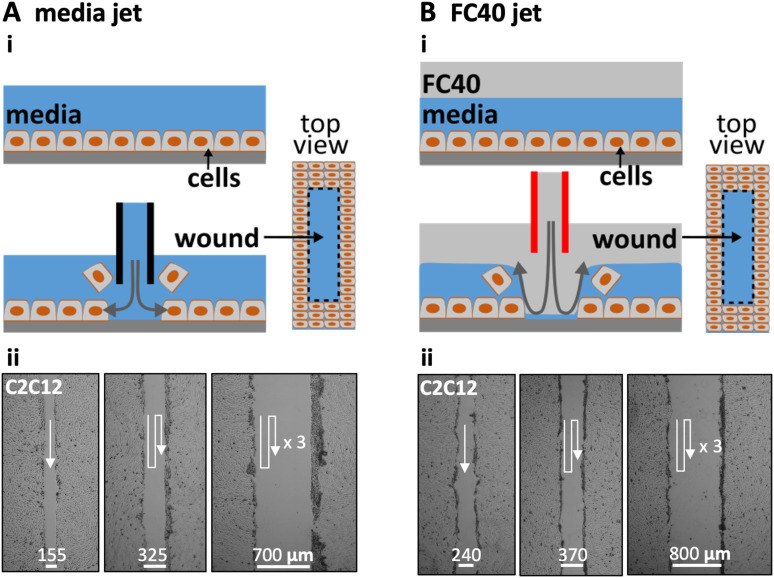
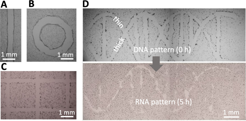
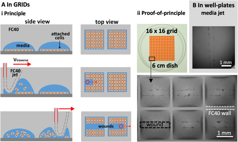
Many wound-healing assays are used in cell biology and biomedicine; they are often labor intensive and/or require specialized and costly equipment. We describe a contactless method to create wounds with any imaginable 2D pattern in cell monolayers using the micro-jets of either media or an immiscible and biocompatible fluorocarbon (i.e., FC40). We also combine this with another method that allows automation and multiplexing using standard Petri dishes. A dish is filled with a thin film of media overlaid with FC40, and the two liquids are reshaped into an array of microchambers within minutes. Each chamber in such a grid is isolated from others by the fluid walls of FC40. Cells are now added, allowed to grow into a monolayer, and wounds are created using the microjets; then, healing is monitored by microscopy. As arrays of chambers can be made using media and Petri dishes familiar to biologists, and as dishes fit seamlessly into their incubators, microscopes, and workflows, we anticipate that this assay will find wide application in wound healing.


Figures

FIG. 1.
FIG. 1.
Creating wounds in a monolayer of mouse C2C12 myoblasts. (a) With media jet. (i) Cartoon showing a monolayer before and after the nozzle of a steel needle filled with media is lowered until just above the cells; on jetting media, the jet momentum detaches some cells from the surface, and moving the nozzle along a predefined path (here into the page) creates a wound (as shown in the top view). (ii) Images of wounds of different widths produced by jetting overlapping lines (white arrows: jetting paths). (b). With FC40 jet. (i) Cartoon showing a monolayer overlaid with FC40 before and after the nozzle of a steel needle filled with FC40 is lowered into the FC40 overlay until just above the media; on jetting FC40, the jet momentum forces the FC40:media interface down so that it plays on the monolayer to dislodge the cells, and moving the nozzle again creates a wound. (ii) Images of wounds with different widths created by jetting overlapping lines (white arrows: jetting paths).
FIG. 2.
FIG. 2.
Effects of varying flow rate (Q˙) and traverse speed (Vtraverse) on wound width and area (Dnozzle= 60 μm, H = 0.4 μm, wound length = 2.5 mm). Scale bars: 200 μm. (a). Varying Q˙ as Vtraverse is held constant at 300 mm/min. (i) For media jets, wound failure occurs at 7.5 μl/s, and increasing the flow produces wider wounds. (ii) For FC40 jets, wound failure occurs at 5.4 μl/s, and increasing the flow again yields wider wounds (often with a central column of FC40 drops that adhere to the dish). (iii) Relationship between (cell-free) wound area and flow rate. Except failure conditions, each datapoint represents an average of at least n = 6 measurements. (b). Varying Vtraverse as Q˙ is held constant at 8 μl/s. (i) For a media jet, increasing the traverse rate leads to wound failure as the dwell time over a given area falls. (ii) For an FC40 jet, failure occurs at a higher traverse rate. (iii) Relationship between the cell-free wound area and the traverse rate. Except failure conditions, each datapoint represents an average of at least n = 6 measurements.
FIG. 3.
FIG. 3.
Wounding patterns. All shapes were created in 6 cm Petri dishes using media jets. (a) Line. (b) Circle. (c) A complex wound (part of 16 × 16 grid). (d) A wound shaped like a double helix of DNA (with one thick backbone strand + attached base and one thin strand + base). After incubation (5 h), the thin strand has almost completely healed, so the structure looks like a single strand of RNA.
FIG. 4.
FIG. 4.
Multiplexed wounding. (a) In grids, with an FC40 jet. (i) Principle. Cells are seeded in chambers in a grid, grown to confluency, and the jet traverses above the centerline of each row; as it passes, the jet forces the FC40:media interface down so that it plays on monolayers to detach the cells. (ii) Image of grid. Chambers initially held 100 nl of media, and 400 nl of red dye is added to aid visualization. Zoom: images of wounds in six chambers. (b). In the well of a conventional 96-well plate, with a media jet.

Similar articles

Cited by

References

    1. Friedl P. and Gilmour D., “Collective cell migration in morphogenesis, regeneration and cancer,” Nat. Rev. Mol. Cell Biol. 10, 445–457 (2009). 10.1038/nrm2720 - DOI - PubMed
    1. Li L., He Y., Zhao M., and Jiang J., “Collective cell migration: Implications for wound healing and cancer invasion,” Burn Trauma 1, 21–26 (2013). 10.4103/2321-3868.113331 - DOI - PMC - PubMed
    1. Grada A., Otero-Vinas M., Prieto-Castrillo F., Obagi Z., and Falanga V., “Research techniques made simple: Analysis of collective cell migration using the wound healing assay,” J. Invest. Dermatol. 137, e11–e16 (2017). 10.1016/j.jid.2016.11.020 - DOI - PubMed
    1. Nakamura M., Dominguez A. N. M., Decker J. R., Hull A. J., Verboon J. M., and Parkhurst S. M., “Into the breach: How cells cope with wounds,” Open Biol. 3, 8 (2018).10.1098/rsob.180135 - DOI - PMC - PubMed
    1. Chouhan D., Dey N., Bhardwaj N., and Mandal B. B., “Emerging and innovative approaches for wound healing and skin regeneration: Current status and advances,” Biomaterials 216, 119267 (2019). 10.1016/j.biomaterials.2019.119267 - DOI - PubMed

LinkOut - more resources