

Anatomical Variation in the Mandibular Foramen in Non-Atrophic and Atrophic Mandibles
- PMID: 33598112
- PMCID: PMC7875102
- DOI: 10.5037/jomr.2020.11404
Anatomical Variation in the Mandibular Foramen in Non-Atrophic and Atrophic Mandibles
Abstract
Objectives: Previous studies of variation in mandibular foramen characteristics with age have involved comparison in different populations, but few data, between non-atrophic and atrophic mandibles are available. The aim of this original article was to compare the position, shape and area of the mandibular foramen between non-atrophic and atrophic mandibles.
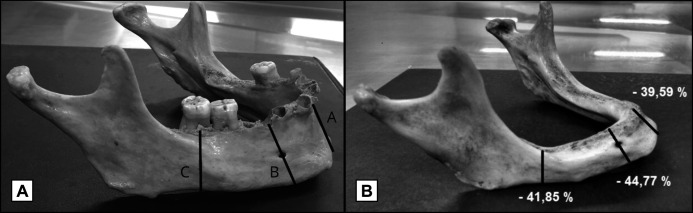
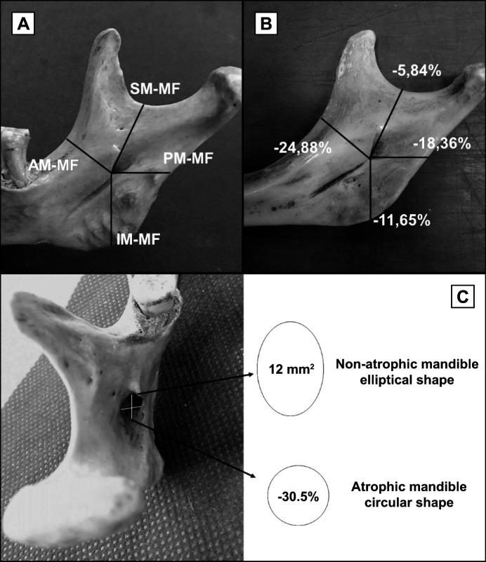
Material and methods: Morphometric methods were used to study the mandibular foramen variation. Fifty adult dry mandibles from the laboratory of anatomy were selected. Mandibles were considered non-atrophic if the distance between the base and alveolar ridge was homogeneous and greater than 25 mm in the anterior region and 20 mm in the posterior region. Conversely, mandibles were considered atrophic if that distances were lower than those described to a minimum of 11 mm in all areas. All measurements were performed with a digital caliper. For statistical analysis, the admitted level of significance was 5%.
Results: When non-atrophic mandibles were compared to atrophic ones, the mandibular foramen shifted significantly to an anterior position (mean difference [MD]: 4.81 mm; P < 0.0001) and to an inferior position (MD: 3.04 mm; P < 0.0001) and changed from an elliptical shape to round one, with a significant decrease in its area (MD: 3.66 mm2; P < 0.05).
Conclusions: The results indicate that there are significant differences in the position, shape and area of the mandibular foramen between non-atrophic and atrophic mandibles. These data should be considered in anaesthetic techniques and surgical procedures to prevent vascular and nervous lesions.
Keywords: anatomy; atrophy; humans; mandible; mental foramen.
Copyright © Mascaro MB, Picoli LC, Reis Matos ST, Sipos Lotaif SC, De Souza MR, Ferreira Calderon M. Published in the JOURNAL OF ORAL & MAXILLOFACIAL RESEARCH (http://www.ejomr.org), 31 December 2020.
Figures
Similar articles
-
The morphometric analysis of the mental foramen in adult dry human mandibles: a study on the South Indian population.J Clin Diagn Res. 2013 Aug;7(8):1547-51. doi: 10.7860/JCDR/2013/6060.3207. Epub 2013 Aug 1. J Clin Diagn Res. 2013. PMID: 24086835 Free PMC article.
-
Accessory Mental Foramina in Dry Mandibles: An Observational Study Along with Systematic Review and Meta-Analysis.Dent J (Basel). 2025 Feb 22;13(3):94. doi: 10.3390/dj13030094. Dent J (Basel). 2025. PMID: 40136722 Free PMC article.
-
Morphometric study on mandibular foramen and incidence of accessory mandibular foramen in mandibles of south Indian population and its clinical implications in inferior alveolar nerve block.Anat Cell Biol. 2016 Dec;49(4):241-248. doi: 10.5115/acb.2016.49.4.241. Epub 2016 Dec 31. Anat Cell Biol. 2016. PMID: 28127498 Free PMC article.
-
The mental foramen and nerve: clinical and anatomical factors related to dental implant placement: a literature review.J Periodontol. 2006 Dec;77(12):1933-43. doi: 10.1902/jop.2006.060197. J Periodontol. 2006. PMID: 17209776 Review.
-
Accessory mental foramen: an anatomical study on dry mandibles and review of the literature.Oral Maxillofac Surg. 2015 Jun;19(2):177-81. doi: 10.1007/s10006-014-0474-1. Epub 2014 Nov 15. Oral Maxillofac Surg. 2015. PMID: 25394607 Review.
Cited by
-
The Influence of Craniometric Variation on the Position of Mandibular Foramen: A Cadaveric Cross-Sectional Study.Medicina (Kaunas). 2024 Nov 23;60(12):1925. doi: 10.3390/medicina60121925. Medicina (Kaunas). 2024. PMID: 39768807 Free PMC article.
References
-
- Kjaer I. Correlated appearance of ossification and nerve tissue in human fetal jaws. J Craniofac Genet Dev Biol. 1990;10(3):329-36. - PubMed
LinkOut - more resources
Full Text Sources