Review
. 2021 Feb 1:8:637838.
doi: 10.3389/fcvm.2021.637838. eCollection 2021.

Noninvasive Imaging in Interventional Cardiology: Hypoplastic Left Heart Syndrome

Affiliations
Review

Noninvasive Imaging in Interventional Cardiology: Hypoplastic Left Heart Syndrome

Hannah Bellsham-Revell. Front Cardiovasc Med. .

Abstract

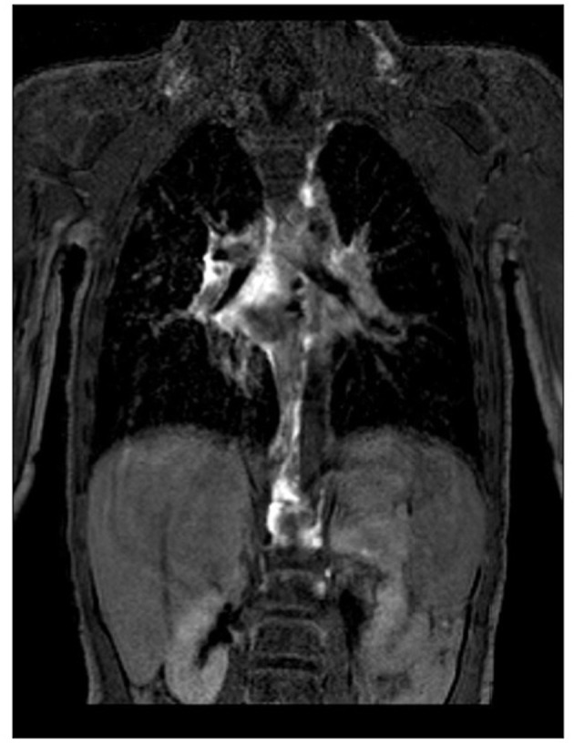
Hypoplastic left heart syndrome (HLHS) is a spectrum of left heart underdevelopment leaving the left side unable to support the systemic circulation. If active management is pursued, then the condition is managed with staged palliation to the Fontan circulation, leaving a systemic right ventricle. Through all surgical stages, and even after completion of Fontan, there are multiple areas that may require intervention, most frequently the branch pulmonary arteries which are essential to a successful Fontan circulation. Echocardiography is the mainstay of assessment, but there is an increasing use of magnetic resonance imaging (MRI) and computed tomography (CT) particularly in relation to extracardiac structures which can be more challenging with echocardiography. Both MRI and CT require set-up, experience and training, and usually sedation or anesthetic in smaller children, but can provide excellent imaging to guide interventions. Cardiac MRI is also able to quantify right ventricular (RV) function which can be challenging on echocardiography. This article describes the modalities available and their use in assessing patients with HLHS prior to catheter interventions.

Keywords: Fontan; Glenn; MRI; Norwood; computed tomography; echocardiography; hybrid; hypoplastic left heart syndrome.


Conflict of interest statement

The author declares that the research was conducted in the absence of any commercial or financial relationships that could be construed as a potential conflict of interest.

Figures

Figure 1
Figure 1
(A) Unoperated hypoplastic left heart syndrome. (B) Classical Norwood procedure. (C) Norwood procedure with Sano modification. (D) Hybrid procedure. (E) Superior cavopulmonary connection with (i) Glenn or (ii) hemi-Fontan. (F) Total cavopulmonary connection with (i) Fenestrated extracardiac conduit or (ii) Fenestrated lateral tunnel.
Figure 2
Figure 2
(A,i) Proximal and (A,ii) distal right ventricle to pulmonary artery conduit on echocardiography. (B) Modified Blalock-Taussig-Thomas shunt on echocardiography. (C,i,ii) Right ventricle to pulmonary artery conduit on CT. (D) Modified Blalock-Taussig-Thomas shunt on CT.
Figure 3
Figure 3
(A) DKS anastomosis on echocardiography. (B) DKS anastomosis on MRI. (C) DKS narrowing on CT. DKS, Damus-Kaye-Stansel; RV, right ventricle.
Figure 4
Figure 4
(A) Arch obstruction on echocardiography shown on 2D and color. (B) Arch obstruction on MRI.
Figure 5
Figure 5
(A) Restrictive atrial communication on color. (B) Elevated transatrial Doppler mean gradient. (C) A wave reversal on pulmonary vein Doppler.
Figure 6
Figure 6
(A) Normal “sawtooth” pulmonary band Doppler pattern. (B) Abnormal “pulsatile” pulmonary band Doppler pattern. (C,i) 3D reconstruction of banded branch pulmonary arteries. (C,ii) Reformat of CT and banded branch pulmonary arteries. LPA, Left pulmonary artery; RPA, right pulmonary artery.
Figure 7
Figure 7
(A) Micro transoesophageal echocardiographic guidance of atrial septal stent. (B) Jailed pulmonary veins seen on echo. (C) CT scan showing atrial septal stent with dilated right pulmonary veins. RA, Right atrium; RPVs, right pulmonary veins.
Figure 8
Figure 8
(A) Narrowing of arterial ductal stent on 2D and color. (B) Increased Doppler gradient on ductal stent. (C) Constriction distal to the ductal stent with involvement of the isthmus and retrograde aortic arch.
Figure 9
Figure 9
(A,i) Right upper pulmonary artery narrowing with Glenn on echocardiography. (A,ii) Proximal left pulmonary artery narrowing – site of previous pulmonary artery band. (B,i) Narrowing of the right pulmonary artery at the bifurcation. (B,ii) Proximal left pulmonary artery narrowing on CT. LPA, Left pulmonary artery; RUPA, right upper pulmonary artery; SVC, superior caval vein.
Figure 10
Figure 10
Narrowing of the Fontan circuit seen on MRI.
Figure 11
Figure 11
(A) 3D echocardiogram showing wire across fenestration. (B) Device in fenestration seen on echocardiography. LAT, Lateral tunnel pouch.
Figure 12
Figure 12
(A) Pulmonary vein stenosis on echocardiography on 2D and color. (B) Abnormal pulmonary vein Doppler due to stenosis. (C) Left pulmonary vein and atrial septal stents on transthoracic echocardiography. (D) Left pulmonary vein and atrial septal stents on CT.
Figure 13
Figure 13
MRI lymphatic imaging.
Figure 14
Figure 14
Thrombus seen in inferior limb of the Fontan circuit at the inferior caval vein (IVC) junction.

References

    1. Leirgul E, Fomina T, Brodwall K, Greve G, Holmstrøm H, Vollset SE, et al. . Birth prevalence of congenital heart defects in Norway 1994-2009—A nationwide study. Am Heart J. (2014) 168:956–64. 10.1016/j.ahj.2014.07.030 - DOI - PubMed
    1. Barron DJ, Kilby MD, Davies B, Wright JGC, Jones TJ, Brawn WJ. Hypoplastic left heart syndrome. Lancet. (2009) 374:551–64. 10.1016/S0140-6736(09)60563-8 - DOI - PubMed
    1. Mascio CE, Irons ML, Ittenbach RF, Gaynor JW, Fuller SM, Kaplinski M, et al. . Thirty years and 1,663 consecutive Norwood procedures: has survival plateaued? J Thorac Cardiovasc Surg. (2019) 158:220–9. 10.1016/j.jtcvs.2018.12.117 - DOI - PubMed
    1. Atz AM, Travison TG, Williams IA, Pearson GD, Laussen PC, Mahle WT, et al. . Prenatal diagnosis and risk factors for preoperative death in neonates with single right ventricle and systemic outflow obstruction: screening data from the Pediatric Heart Network Single Ventricle Reconstruction Trial(*). J Thorac Cardiovasc Surg. (2010) 140:1245–50. 10.1016/j.jtcvs.2010.05.022 - DOI - PMC - PubMed
    1. Akintürk H, Michel-Behnke I, Valeske K, Mueller M, Thul J, Bauer J, et al. . Hybrid transcatheter–surgical palliation. Pediatr Cardiol. (2007) 28:79–87. 10.1007/s00246-006-1444-7 - DOI - PubMed

LinkOut - more resources