Case Reports
. 2021 Feb 1:8:584975.
doi: 10.3389/fvets.2021.584975. eCollection 2021.

Case Report: Surgical Correction of a Cystic Duct Stump Leakage Following Cholecystectomy Using an Autologous Rectus Sheath Graft in a Dog

Affiliations
Case Reports

Case Report: Surgical Correction of a Cystic Duct Stump Leakage Following Cholecystectomy Using an Autologous Rectus Sheath Graft in a Dog

Hyun-Jung Han et al. Front Vet Sci. .

Abstract

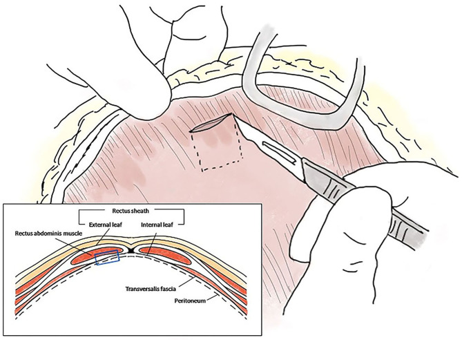
A 2.7 kg, 13-year-old, castrated male Yorkshire Terrier was presented with bile peritonitis after cholecystectomy. Exploratory coeliotomy to identify and correct bile leakage revealed that the transected end of the cystic duct was open with no in-situ ligatures or vascular clips. The residual cystic duct stump was too short to ligate or seal directly. An autologous rectus sheath graft, harvested from the internal leaf of the rectus sheath, was used to patch the cystic duct stump. The graft was secured over the open duct using several simple interrupted sutures and covered with an omentalization. The clinical signs resolved after surgery, except for a transient increase in hepatobiliary enzyme levels and intrahepatic bile duct dilatation. The enzyme levels returned to near normal on day 25 after surgery. No intrahepatic bile duct dilatation was detected on day 55 after surgery. The owner was contacted for 3 years post-operatively and reported that the dog remained healthy without any long-term complications. Grafting using autologous rectus sheath can be used to treat cystic duct stump leakage that cannot be managed with direct closure using traditional modalities due to spatial constraints.

Keywords: autologous rectus sheath; bile peritonitis; cholecystectomy; cystic duct stump leakage; graft; yorkshire terrier.


Conflict of interest statement

The authors declare that the research was conducted in the absence of any commercial or financial relationships that could be construed as a potential conflict of interest.

Figures

Figure 1
Figure 1
Closure of the cystic duct stump using an autologous rectus sheath graft in a Yorkshire Terrier. Images (A,C,E) are intaoperative photographs; (B,D,F) incorporate schematic overlays to highlight the relevant anatomy and location of suture placement. (A,B) The transected end of the cystic duct is completely open (white circle) and is close to the base of the bilateral hepatic ducts and the common bile duct (green color plane with black solid line). The white semi-transparent plane with black dotted line indicates the location of the removed gall bladder and cystic duct. (C,D) The 1 × 1 cm rectus sheath graft (yellow plane) is sutured to the end of the cystic duct stump with simple interrupted sutures using 5–0 polyglyconate (black × marks). The abundant dorsal part of the sheath is sutured to the fibrous capsule of the surrounding right medial liver lobe (white × marks). (E,F) After confirmation of complete closure of the cystic duct, greater omentum is tacked over the patched cystic duct stump by suturing with the fibrous capsule of the right medial liver lobe (blue plane). RML, right medial lobe; RLL, left lateral lobe; QL, quadrate lobe; ST, stomach.
Figure 2
Figure 2
Schematic diagram of the autologous rectus sheath graft. The internal leaf of the rectus sheath was approached via the coeliotomy site, and a 1 × 1 cm sheath with peritoneum was peeled off at the level of the midline, ~1 cm from the incision (blue empty square in the box).
Figure 3
Figure 3
Post-operative ultrasound images of the hepatic parenchyma of a Yorkshire Terrier with unusual cystic duct stump leakage treated using an autologous rectus sheath graft. Color Doppler imaging shows intrahepatic bile duct dilatation in images (A,B) (white arrow). The intrahepatic bile duct diameter is 0.7 mm on post-operative day 5 (A), 1.4 mm on post-operative day 25 (B), and not measurable on post-operative day 55 (C).

Similar articles

Cited by

References

    1. Mehler SJ, Mayhew PD, Drobatz KJ, Holt DE. Variables associated with outcome in dogs undergoing extrahepatic biliary surgery: 60 cases (1988–2002). Vet Surg. (2004) 33:644–9. 10.1111/j.1532-950X.2004.04087.x - DOI - PubMed
    1. Mehler SJ. Complications of the extrahepatic biliary surgery in companion animals. Vet Clin North Am Small Anim Pract. (2011) 41:949–67. 10.1016/j.cvsm.2011.05.009 - DOI - PubMed
    1. Woods MS, Shellito JL, Santoscoy GS, Hagan RC. Cystic duct leaks in laparoscopic cholecystectomy. Am J Surg. (1994) 168:560–5. 10.1016/S0002-9610(05)80122-9 - DOI - PubMed
    1. Eisenstein S, Greenstein AJ, Kim U, Divino CM. Cystic duct stump leaks: after the learning curve. Arch Surg. (2008) 143:1178–83. 10.1001/archsurg.143.12.1178 - DOI - PubMed
    1. Pike FS, Berg J, King NW, Penninck DG, Webster CR. Gallbladder mucocele in dogs: 30 cases (2000–2002). J Am VetMed Assoc. (2004) 224:1615–22. 10.2460/javma.2004.224.1615 - DOI - PubMed

Publication types

LinkOut - more resources