Review
. 2021 May 7;104(5):976-994.
doi: 10.1093/biolre/ioab023.

Role of zinc in female reproduction

Affiliations
Review

Role of zinc in female reproduction

Tyler Bruce Garner et al. Biol Reprod. .

Abstract

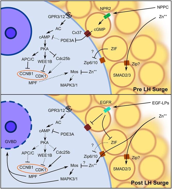
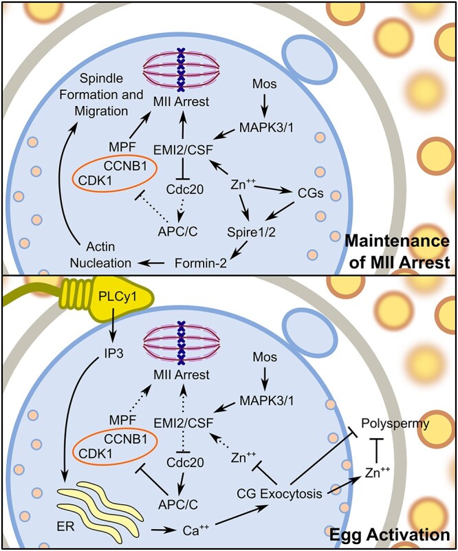
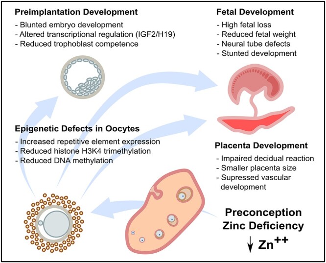
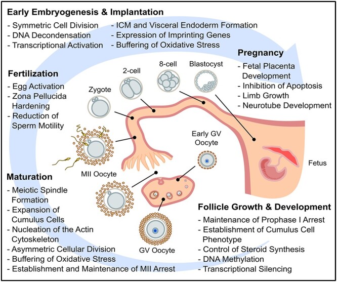
Zinc is a critical component in a number of conserved processes that regulate female germ cell growth, fertility, and pregnancy. During follicle development, a sufficient intracellular concentration of zinc in the oocyte maintains meiotic arrest at prophase I until the germ cell is ready to undergo maturation. An adequate supply of zinc is necessary for the oocyte to form a fertilization-competent egg as dietary zinc deficiency or chelation of zinc disrupts maturation and reduces the oocyte quality. Following sperm fusion to the egg to initiate the acrosomal reaction, a quick release of zinc, known as the zinc spark, induces egg activation in addition to facilitating zona pellucida hardening and reducing sperm motility to prevent polyspermy. Symmetric division, proliferation, and differentiation of the preimplantation embryo rely on zinc availability, both during the oocyte development and post-fertilization. Further, the fetal contribution to the placenta, fetal limb growth, and neural tube development are hindered in females challenged with zinc deficiency during pregnancy. In this review, we discuss the role of zinc in germ cell development, fertilization, and pregnancy with a focus on recent studies in mammalian females. We further detail the fundamental zinc-mediated reproductive processes that have only been explored in non-mammalian species and speculate on the role of zinc in similar mechanisms of female mammals. The evidence collected over the last decade highlights the necessity of zinc for normal fertility and healthy pregnancy outcomes, which suggests zinc supplementation should be considered for reproductive age women at risk of zinc deficiency.

Keywords: female; mammal; zinc.


Figures

Figure 1
Figure 1
Diagram of the signaling pathways that regulate GV arrest and GVBD. The NPPC secreted from peripheral MGCs binds to NPR2 in CGCs to upregulate cGMP synthesis. The cGMP passively diffuses into the GV oocyte via Cx37 predominant gap junctions to inhibit PDE3A, thus maintaining an elevated concentration of cAMP in the oocyte through constitutive activation of AC by GPR3/12. The high concentration of cAMP activates PKA to directly inhibit MPF through APC/C proteasomal degradation of CCNB1 and WEE1B phosphorylation of CDK1 in addition to inhibiting Cdc25b activity. The CGCs also secrete ZIF that prevents uptake of free intracellular zinc by the oocyte, possibly through inhibition of Zip6/10. In the CGCs, zinc is required for SMAD2/3 activation to maintain the cumulus phenotype, while in GV oocytes, zinc inhibits premature activation of the Mos–MAPK3/1 pathway. Following the LH surge, EGF-LPs secreted by MGCs bind to the EGFR in CGCs to initiate the closure of CGC–oocyte gap junctions, thus preventing further cGMP diffusion into the oocyte. Activation of the EGFR in CGCs also inhibits ZIF activity, allowing free intracellular zinc accumulation in the oocyte during maturation. The reduction in intra-oocyte cGMP concentration increases PDE3A activity to degrade cAMP. As PKA activity is subsequently reduced, MPF is activated by increased CCNB1 availability and CDK1 dephosphorylation by Cdc25b, leading to both activation of Mos–MAPK3/1 and GVBD. Abbreviations: AC, adenylyl cyclase; EGFR, epidermal growth factor receptor; Zn++, zinc. Arrows indicate activation and bars represent inhibition. Solid lines indicate active pathways, while dashed lines represent inactive pathways.
Figure 2
Figure 2
Model of signaling pathways that modulate MII arrest and egg activation. A high intracellular concentration of zinc establishes and maintains MII arrest through activation of EMI2 to prevent degradation of CCNB1 by the APC/C proteasome and maintains high MPF activity to maintain MII arrest. Additionally, zinc accumulation facilitates Spire1/2 localization with Formin-2 to nucleate actin cytoskeleton and to facilitate meiotic spindle formation and migration to the cortex. Following sperm binding and the acrosomal reaction, PLCy1 increases IP3 availability that initiates calcium release from the ER. This calcium influx promotes exocytosis of CGs, releasing zinc and other factors important for establishing the block to polyspermy. The subsequent reduced intracellular concentration of zinc reduces EMI2 activity. Subsequent activation of the APC/C proteasome by Cdc20 leads to the degradation of CCNB1 to reduce MPF activity, thus allowing completion of meiosis. Abbreviations: Ca++, calcium; ER, endoplasmic reticulum; Zn++, zinc. Arrows indicate activation and bars represent inhibition. Solid lines indicate active pathways, while dashed lines represent inactive pathways.
Figure 3
Figure 3
Epigenetic defects caused by preconception ZD. A 3–5 day period of dietary ZD is sufficient to cause a reduction in DNA and histone methylation and increased abundance of transcripts from the repetitive elements in the oocyte. These defects are associated with impaired preimplantation development, including reduced abundance of Igf2/H19 transcripts and impaired trophoblast differentiation. However, even later in pregnancy after normal dietary zinc status is restored, defects in placenta formation, fetal survival, and fetal development persist, suggesting a long-lasting impairment of developmental potential.
Figure 4
Figure 4
Summary of the events and processes in mammalian female reproduction that zinc is known to play a role.

References

    1. Swenerton H, Shrader R, Hurley LS. Zinc-deficient embryos: Reduced thymidine incorporation. Science 1969; 166:1014–1015. - PubMed
    1. Duncan JR, Hurley LS. Thymidine kinase and DNA polymerase activity in normal and zinc deficient developing rat embryos. Proc Soc Exp Biol Med 1978; 159:39–43. - PubMed
    1. Chanfreau GF. Zinc’ing down RNA polymerase I. Transcription 2013; 4:217–220. - PMC - PubMed
    1. Maret W. Zinc biochemistry: From a single zinc enzyme to a key element of life. Adv Nutr 2013; 4:82–91. - PMC - PubMed
    1. Wallwork JC, Duerre JA. Effect of zinc deficiency on methionine metabolism, methylation reactions and protein synthesis in isolated perfused rat liver. J Nutr 1985; 115:252–262. - PubMed

Publication types