Review
. 2021 Sep;50(9):1763-1773.
doi: 10.1007/s00256-021-03734-7. Epub 2021 Feb 18.

Musculoskeletal involvement of COVID-19: review of imaging

Affiliations
Review

Musculoskeletal involvement of COVID-19: review of imaging

Santhoshini Leela Ramani et al. Skeletal Radiol. 2021 Sep.

Abstract

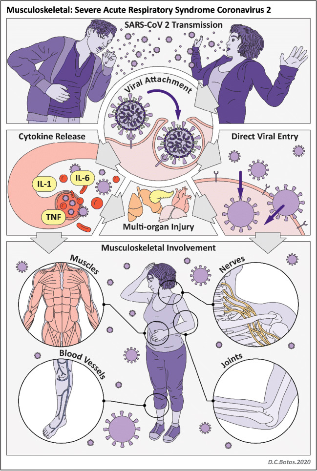
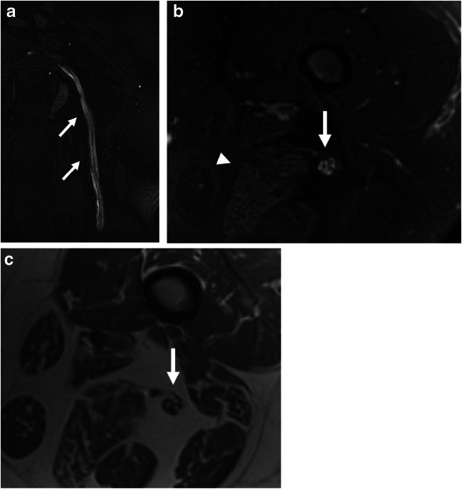
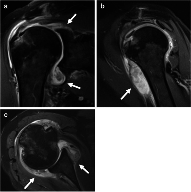
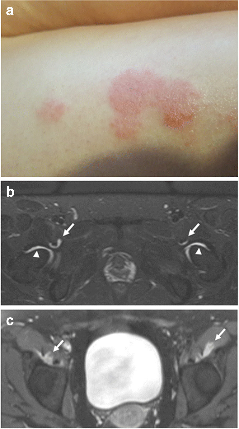
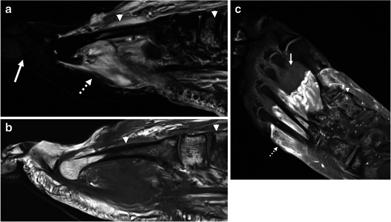
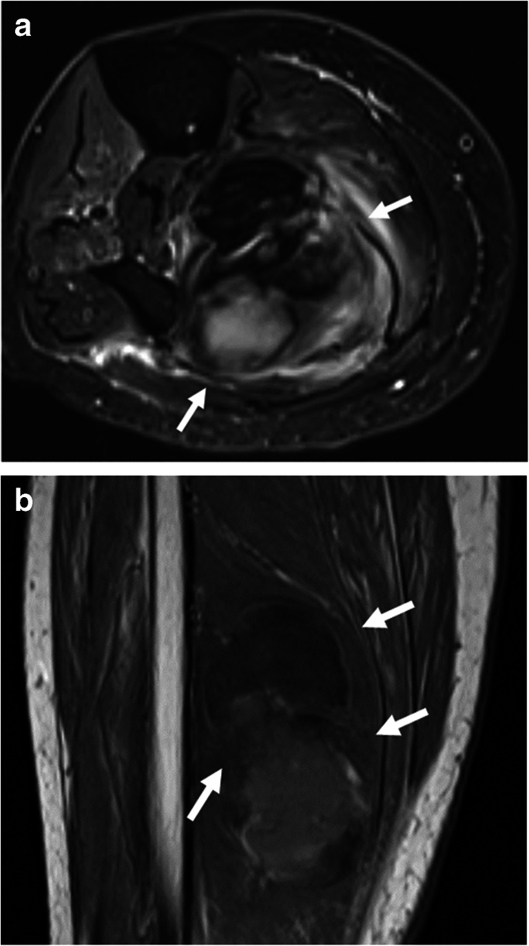
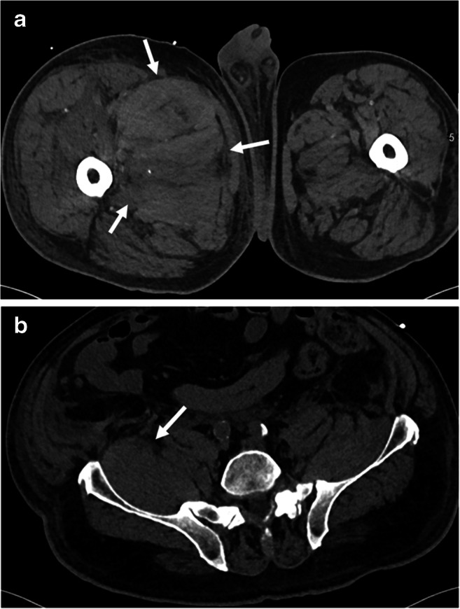
The global pandemic of coronavirus disease 2019 (COVID-19) has revealed a surprising number of extra-pulmonary manifestations of severe acute respiratory syndrome coronavirus 2 (SARS-CoV-2) infection. While myalgia is a common clinical feature of COVID-19, other musculoskeletal manifestations of COVID-19 were infrequently described early during the pandemic. There have been emerging reports, however, of an array of neuromuscular and rheumatologic complications related to COVID-19 infection and disease course including myositis, neuropathy, arthropathy, and soft tissue abnormalities. Multimodality imaging supports diagnosis and evaluation of musculoskeletal disorders in COVID-19 patients. This article aims to provide a first comprehensive summary of musculoskeletal manifestations of COVID-19 with review of imaging.

Keywords: COVID-19; MR; Musculoskeletal; Neuromuscular; SARS-CoV-2; Ultrasound.


Conflict of interest statement

The authors declare no competing interests.

Figures

Fig. 1
Fig. 1
Illustration of SARS-CoV-2 infection
Fig. 2
Fig. 2
A 44-year-old male with incidental positive COVID-19 test obtained prior to eye surgery. He did not have any respiratory symptoms and was surprised by the positive test. In the following 2–3 weeks, he developed progressive weakness and swelling requiring hospitalization. MR imaging of the upper and lower extremities with intravenous contrast was performed. a Axial T2-weighted fat-saturated and b axial post-contrast T1-weighted fat-saturated images demonstrate diffuse edema and enhancement of the proximal right upper extremity musculature (arrows). c Coronal T2-weighted fat-saturated image of the left lower extremity demonstrates muscle edema of the proximal muscles/limb girdle (arrow) with sparing of the distal muscles. Subcutaneous soft tissue edema is noted. Biopsy of the deltoid muscle was performed. d Histopathologic findings included scattered pale degenerating myofibers, surrounded by macrophages (arrow) on H&E stain. e Acid phosphatase highlighted the macrophages (arrow). f NADH stain showed an unusual ring-like pattern to the myofibrillar architecture (scale bar = 100 μm). The patient was diagnosed with post-infectious inflammatory necrotizing myositis and discharged on oral prednisone with subsequent clinical improvement and decrease in CK levels on follow-up
Fig. 3
Fig. 3
A 53-year-old male with prolonged hospitalization for COVID-19 ARDS who developed multiple peripheral nerve injuries during his hospital course and presented with clinical concern of right phrenic nerve palsy. a Ultrasound of the right hemidiaphragm (arrowheads) at expiration and b inspiration demonstrates little change in thickness of the hemidiaphragm muscle, compatible with decreased contractility function. There is no evidence of right hemidiaphragm muscle atrophy. c High-resolution ultrasound of the phrenic nerve (arrow, calipers) demonstrates normal size and echogenicity. Differential diagnosis for hemidiaphragm paralysis includes critical illness myopathy, ventilator-induced diaphragm dysfunction, and the hypothetical plausibility of virus-related myopathy
Fig. 4
Fig. 4
A 60-year-old male with prolonged hospitalization for COVID-19 ARDS including prone positioning who developed multiple peripheral nerve injuries during his hospital course and presented with severe neck and shoulder pain. Clinical concern was for right spinal accessory nerve injury. a Longitudinal and b transverse high-resolution ultrasound demonstrates thickening and hypoechogenicity of the right spinal accessory nerve (arrows). c Longitudinal and d transverse high-resolution ultrasound of the normal left spinal accessory nerve (arrows) is shown for comparison
Fig. 5
Fig. 5
A 37-year-old female with history of hospitalization for COVID-19 complicated by sacral ulcers presenting with left sciatic mononeuropathy. a Coronal post-contrast (for vascular suppression) T2 SPACE and b axial T2-weighted fat-saturated images of the left femur demonstrate diffuse signal hyperintensity of the sciatic nerve (arrow). Subtle asymmetric fatty atrophy and edema of the posterior muscle compartment of the thigh (arrowhead) is suggestive of early denervation. c Axial T1-weighted anatomic image identifies the sciatic nerve (arrow). Based on MR imaging, she was referred to a neuromuscular specialist whose clinical evaluation supported the diagnosis of compressive sciatic neuropathy acquired during her critical illness
Fig. 6
Fig. 6
A 72-year-old female with history of rheumatoid arthritis that had been dormant for over 2 years who experienced multi-joint rheumatoid arthritis flair after contracting COVID-19 (with documented positive testing). Of note, she experienced only mild symptoms with her acute SARS-CoV-2 infection. a Coronal, b sagittal, and c axial post-contrast T1-weighted images of the right shoulder demonstrate moderate enhancing synovitis with extensive low signal intensity debris (arrows). Her symptoms improved following intra-articular steroid injection
Fig. 7
Fig. 7
A 30-year-old female with new-onset skin rashes and multiple arthralgias approximately 2 weeks after COVID-19. a Physical exam showed papules and plaques with areas of erythema, scaling, and lichenification over the extremities, axillae, and abdomen. Skin biopsy confirmed diagnosis of psoriasis. b Axial STIR and c post-contrast T1-weighted fat-saturated images of the pelvis demonstrate mild bilateral hip synovitis (arrowheads) and iliopsoas bursitis (arrows). She was diagnosed with COVID-19-triggered psoriatic arthritis and treated with methotrexate, non-steroidal anti-inflammatory drugs, and corticosteroids
Fig. 8
Fig. 8
A 57-year-old male with history of prostate cancer and prolonged hospitalization for COVID-19 with multi-organ failure requiring vasopressors. a Sagittal STIR image demonstrates soft tissue edema of the proximal foot (dashed arrow) but no edema distally (solid arrow). Heterogeneous bone marrow edema pattern (arrowhead) is seen on both STIR and b sagittal T1-weighted image. c Post-contrast T1-weighted fat-saturated image demonstrates soft tissue enhancement of the proximal foot (dashed arrow) with non-enhancing devitalized tissue distally (solid arrow), compatible with gangrene. Multifocal bone marrow edema pattern (arrowhead) is compatible with associated osteonecrosis
Fig. 9
Fig. 9
A 71-year-old female with COVID-19 ARDS complicated by stroke and deep venous thrombosis, treated with systemic anticoagulation. She developed mass-like swelling of the leg. a Axial T2-weighted fat-saturated and b coronal T1-weighted images demonstrate an intramuscular hematoma (arrows)
Fig. 10
Fig. 10
A 76-year-old male with COVID-19 on therapeutic anti-coagulation who developed right femoral and obturator neuropathies. a, b Axial CT demonstrates a large hematoma of the proximal right thigh musculature extending into the retroperitoneum along the right iliopsoas muscle (arrows)

Similar articles

Cited by

References

    1. Johns Hopkins University & Medicine Coronavirus Resource Center. https://coronavirus.jhu.edu/map.html. Accessed 1/10/21
    1. Revzin MV, Raza S, Warshawsky R, et al. Multisystem imaging manifestations of COVID-19. Part 1. Viral pathogenesis and pulmonary and vascular system complications. Radiographics. 2020;40(6):1574–1599. doi: 10.1148/rg.2020200149. - DOI - PMC - PubMed
    1. Revzin MV, Raza S, Srivastava NC, et al. Multisystem imaging manifestations of COVID-19. Part 2. From cardiac complications to pediatric manifestations. Radiographics. 2020;40(7):1866–1892. doi: 10.1148/rg.2020200195. - DOI - PMC - PubMed
    1. Wiersinga WJ, Rhodes A, Cheng AC, Peacock SJ, Prescott HC. Pathophysiology, transmission, diagnosis, and treatment of coronavirus disease 2019 (COVID-19): a review. JAMA. 2020;324(8):782–793. doi: 10.1001/jama.2020.12839. - DOI - PubMed
    1. Paliwal VK, Garg RK, Gupta A, Tejan N. Neuromuscular presentations in patients with COVID-19. Neurol Sci. 2020;41(11):3039–3056. doi: 10.1007/s10072-020-04708-8. - DOI - PMC - PubMed