

Discrimination of myogenic and nonmyogenic cells from embryonic skeletal muscle by 90 degrees light scattering
- PMID: 3359891
- PMCID: PMC4128021
- DOI: 10.1002/cyto.990090204
Discrimination of myogenic and nonmyogenic cells from embryonic skeletal muscle by 90 degrees light scattering
Abstract
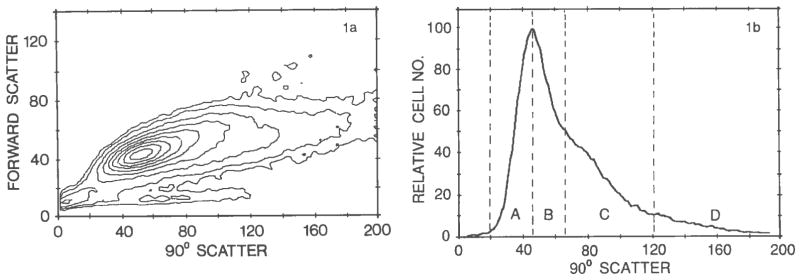
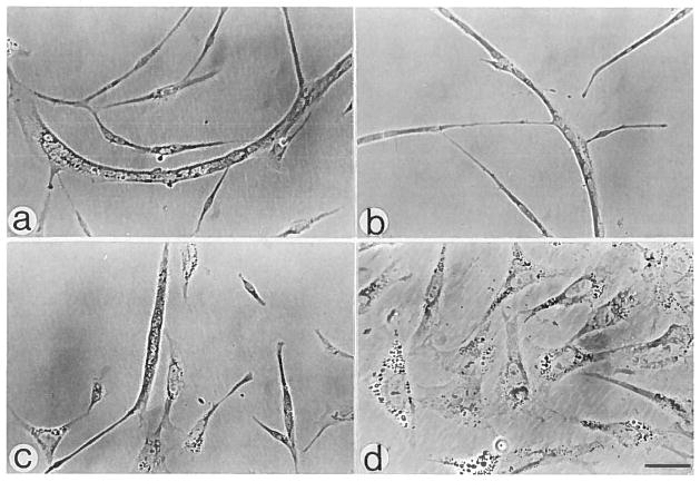
A myogenic cell suspension was isolated from the breast muscles of 10-day-old chicken embryos by trypsin digestion. The cell preparation was subjected to Percoll density centrifugation to reduce the number of fibroblast-like cells present. The Percoll-isolated, predominantly myogenic cell population was then fractionated by flow cytometry using 90 degrees light scattering as the parameter for sorting. Cells exhibiting lower scatter, with a peak of 45 units, produced cultures containing myotubes and gave rise only to myogenic clones. Cells exhibiting higher scatter (120-200 units) produced nonmyogenic cultures and gave rise to nonmyogenic clones. Cells with intermediate light scatter were also detected. The latter produced both myogenic and nonmyogenic clones. The differences in light scatter presumably reflect higher cytoplasmic complexity of the nonmyogenic cells compared with the myogenic cells. Moreover, the differences in light scattering properties of the different cell types offer a means for the isolation of pure populations of myogenic cells directly from the intact muscle.
Figures
Similar articles
-
Skeletal muscle cell populations. Separation and partial characterization of fibroblast-like cells from embryonic tissue using density centrifugation.Histochemistry. 1987;87(1):27-38. doi: 10.1007/BF00518721. Histochemistry. 1987. PMID: 3038797 Free PMC article.
-
Isolation and clonal analysis of satellite cells from chicken pectoralis muscle.Dev Biol. 1987 Jan;119(1):252-9. doi: 10.1016/0012-1606(87)90226-0. Dev Biol. 1987. PMID: 3025033 Free PMC article.
-
Separation of functionally divergent muscle precursor cell populations from porcine juvenile muscles by discontinuous Percoll density gradient centrifugation.BMC Cell Biol. 2018 Mar 9;19(1):2. doi: 10.1186/s12860-018-0156-1. BMC Cell Biol. 2018. PMID: 29523096 Free PMC article.
-
Selection of defined cell types by flow-cytometric cell sorting.Trends Biotechnol. 1993 Feb;11(2):55-62. doi: 10.1016/0167-7799(93)90123-Q. Trends Biotechnol. 1993. PMID: 7763479 Review.
-
Light scatter of isolated cell nuclei as a parameter discriminating the cell-cycle subcompartments.Methods Cell Biol. 1994;41:389-400. doi: 10.1016/s0091-679x(08)61730-6. Methods Cell Biol. 1994. PMID: 7861971 Review. No abstract available.
Cited by
-
Skeletal muscle satellite cells: background and methods for isolation and analysis in a primary culture system.Methods Mol Biol. 2012;798:21-52. doi: 10.1007/978-1-61779-343-1_2. Methods Mol Biol. 2012. PMID: 22130829 Free PMC article.
-
Reporter-Based Isolation of Developmental Myogenic Progenitors.Front Physiol. 2018 Apr 5;9:352. doi: 10.3389/fphys.2018.00352. eCollection 2018. Front Physiol. 2018. PMID: 29674978 Free PMC article.
-
The skeletal muscle satellite cell: still young and fascinating at 50.J Histochem Cytochem. 2011 Dec;59(12):1041-59. doi: 10.1369/0022155411426780. J Histochem Cytochem. 2011. PMID: 22147605 Free PMC article.
-
Extraocular muscle satellite cells are high performance myo-engines retaining efficient regenerative capacity in dystrophin deficiency.Dev Biol. 2015 Jan 1;397(1):31-44. doi: 10.1016/j.ydbio.2014.08.035. Epub 2014 Sep 16. Dev Biol. 2015. PMID: 25236433 Free PMC article.
References
-
- Bailey AJ, Shellswell GB, Duance VC. Identification and change of collagen types in differentiating myoblasts and developing chick muscle. Nature. 1979;278:67–69. - PubMed
-
- Garrett DM, Conrad GW. Fibroblast-like cells from embryonic chick cornea, heart and skin are antigenically distinct. Dev Biol. 1979;70:50–70. - PubMed
-
- Hauschka SD. Clonal analysis of vertebrate myogenesis. III. Developmental changes in the muscle-colony-forming cells of the human fetal limb. Dev Biol. 1974;37:345–368. - PubMed
Publication types
MeSH terms
Grants and funding
LinkOut - more resources
Full Text Sources