. 2021 Aug 1;529(11):2842-2864.
doi: 10.1002/cne.25130. Epub 2021 Feb 28.

Macaque monkey trigeminal blink reflex circuits targeting orbicularis oculi motoneurons

Affiliations

Macaque monkey trigeminal blink reflex circuits targeting orbicularis oculi motoneurons

Paul J May et al. J Comp Neurol. .

Abstract

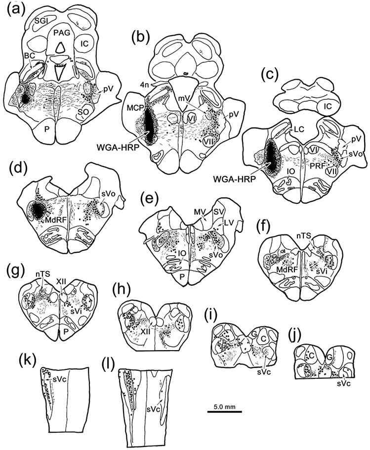
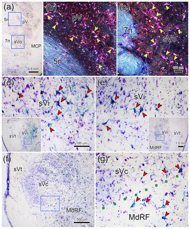
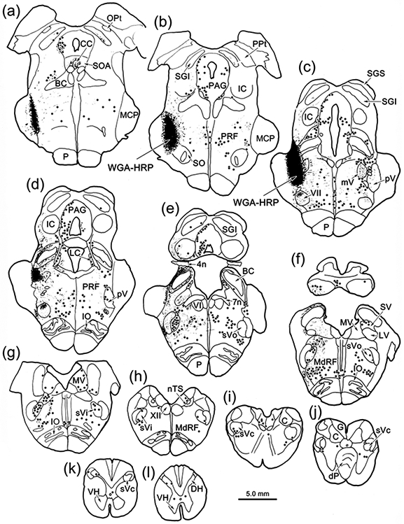
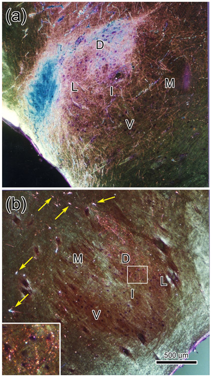
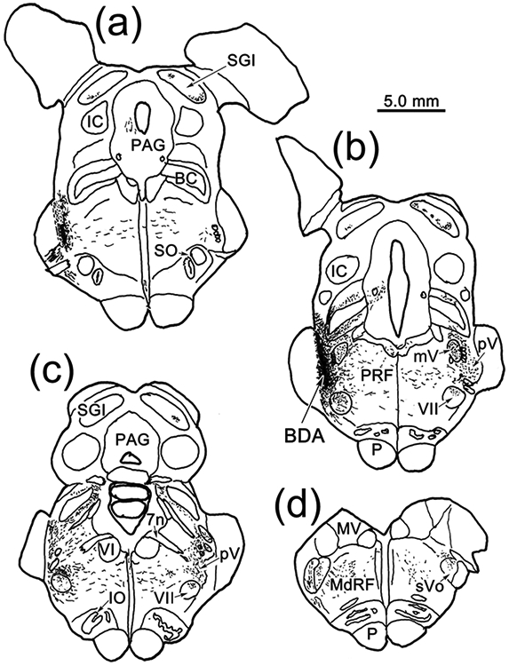
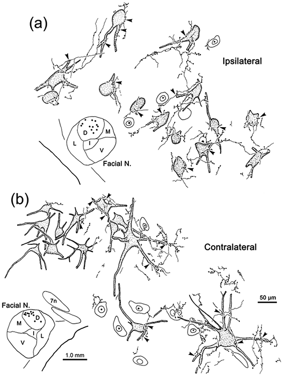
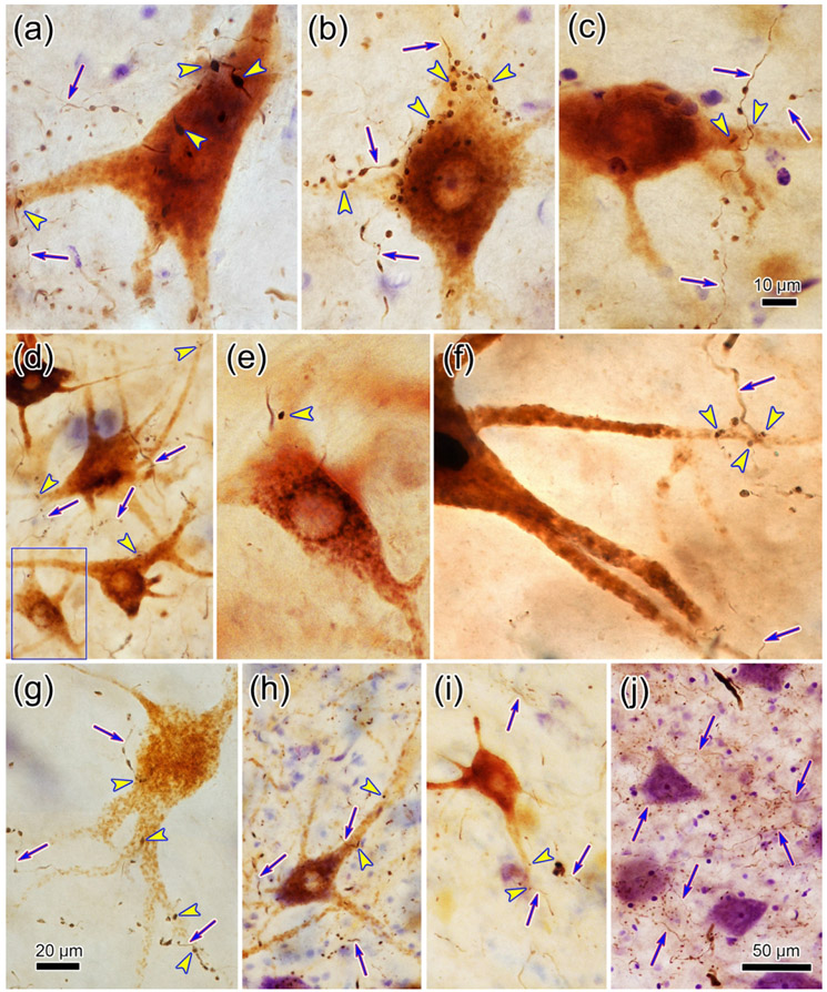
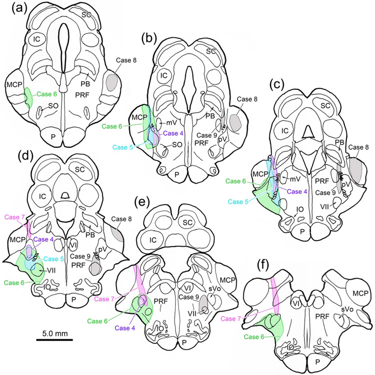
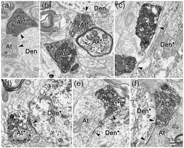
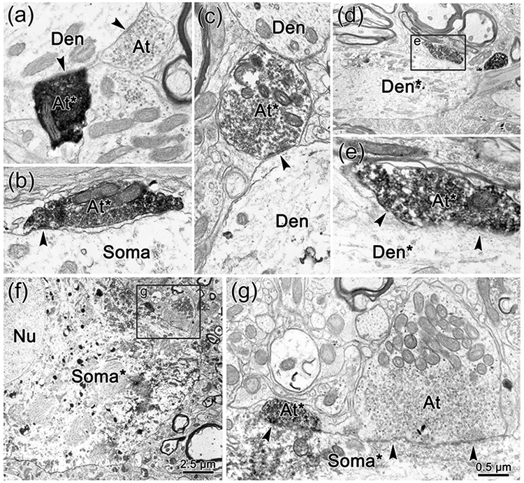
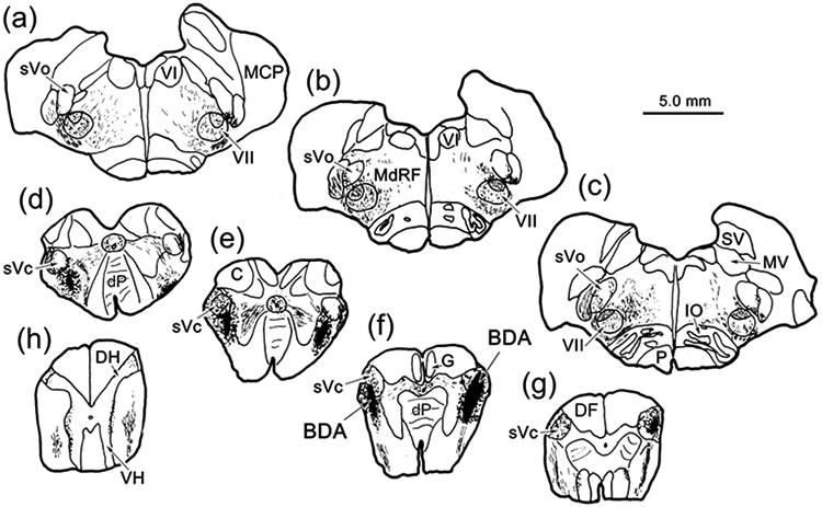
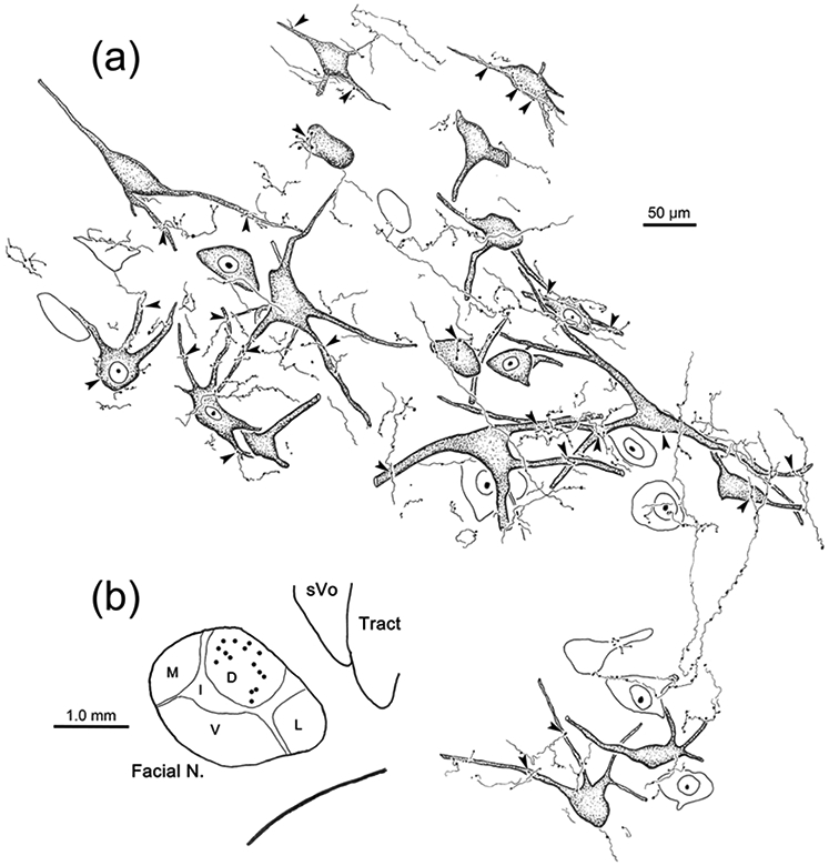
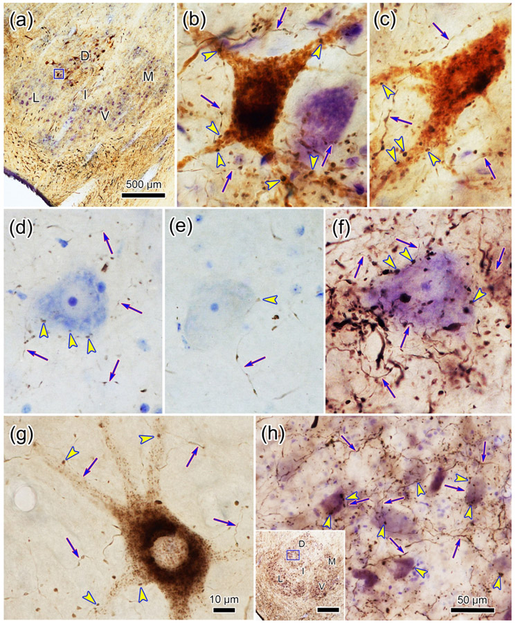
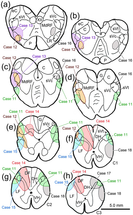
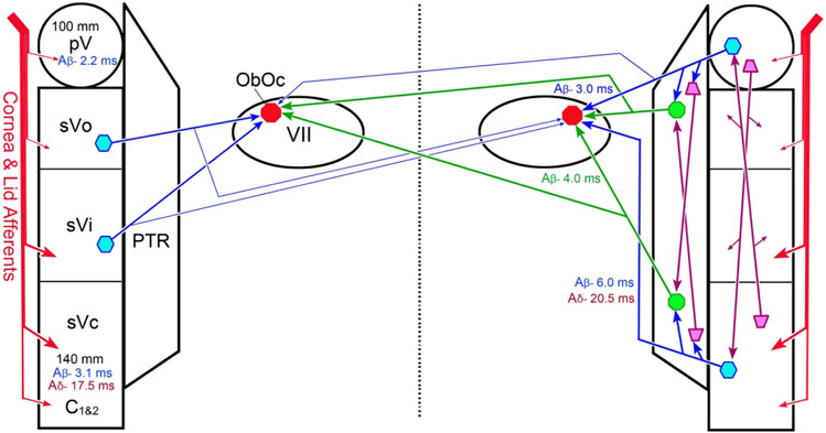
The trigeminal blink reflex plays an important role in protecting the corneal surface from damage and preserving visual function in an unpredictable environment. The closing phase of the human reflex, produced by activation of the orbicularis oculi (ObOc) muscles, consists of an initial, small, ipsilateral R1 component, followed by a larger, bilateral R2 component. We investigated the circuitry that underlies this reflex in macaque (Macaca fascicularis and Macaca mulatta) monkeys by the use of single and dual tracer methods. Injection of retrograde tracer into the facial nucleus labeled neurons in the principal trigeminal nucleus, and in the spinal nucleus pars oralis and interpolaris, bilaterally, and in pars caudalis, ipsilaterally. Injection of anterograde tracer into the principal trigeminal nucleus labeled axons that directly terminated on ObOc motoneurons, with an ipsilateral predominance. Injection of anterograde tracer into pars caudalis of the spinal trigeminal nucleus labeled axons that directly terminated on ipsilateral ObOc motoneurons. The observed pattern of labeling indicates that the reticular formation ventromedial to the principal and spinal nuclei also contributes extensive bilateral input to ObOc motoneurons. Thus, much of the trigeminal sensory complex is in a position to supply a monosynaptic drive for lid closure, and the adjacent reticular formation can supply a disynaptic drive. These findings indicate that the assignment of the R1 and R2 components of the blink reflex to different parts of the trigeminal sensory complex cannot be exclusively based on subdivision connectional relationships with facial motoneurons. The characteristics of the R2 component may be due, instead, to other circuit properties.

Keywords: associative learning; classical conditioning; eyelid; facial nucleus; primate; somatosensory; trigeminal nucleus.


Conflict of interest statement

Conflict of Interest: Neither author has any conflicts of interest, financial or otherwise, with respect to the work described in this manuscript.

Figures

Figure 1.
Figure 1.
Brainstem distribution of cells supplying input to VII. An injection of WGA-HRP that included VII (b-c), as well as pV (a-b) and sVo (c-d), retrogradely labeled neurons (dots), anterogradely labeled terminals (stipple), and labeled axons (lines) throughout the brainstem (a-j) and rostral levels of the cervical spinal cord (k-l). Examples a-j are frontal sections arranged in rostral to caudal order, as are the chartings in other figures. The rostral spinal cord (k-l) was cut horizontally.
Figure 2.
Figure 2.
Retrogradely labeled neurons (arrowheads) from the injection shown in figure 1.a-c: Numerous retrogradely labeled neurons are present contralateral to the injection in pV (b) and sVo (c) of the pons, as demonstrated with crossed polarizers. The upper and lower boxes in a indicate the regions shown in b and c, respectively. d-e: The ventral portion of sVi, the location of the ophthalmic subdivision, contains numerous retrogradely labeled neurons on the ipsilateral (d) and contralateral (e) sides. Boxes in the inserts indicate the location of samples d and e. f-g: Retrogradely labeled neurons were also present in the ophthalmic portion of sVc, ipsilaterally. However, even more retrogradely labeled neurons were present in the adjacent medullary reticular formation (MdRF). Box in f indicates region shown ingG. Dots indicate the border between sVc and MdRF in g. Scale in c = b; in d = e&g; and in insert e = insert d.
Figure 3.
Figure 3.
Brainstem distribution of pV connections. An injection of WGA-HRP that included pV (c), retrogradely labeled neurons (dots), anterogradely labeled terminals (stipple), and labeled axons (lines) throughout the brainstem (a-j) and in rostral levels of the cervical spinal cord (k-l). Note the presence of dense terminal labeling in ipsilateral VII (c-d).
Figure 4.
Figure 4.
Trigeminal terminal fields in VII under crossed polarizer illumination. Following a WGA-HRP injection that included pV and rostral sVo (see Fig. 3), dense anterogradely labeled terminal fields were present in the dorsal (D), lateral (L) and intermediate (I) subdivisions of ipsilateral VII (a), with far fewer labeled terminals present in the ventral (V) and medial (M) subdivisions. On the contralateral side (b) only a very sparse projection to the dorsal division was present. The portion of the contralateral terminal field in the boxed region is shown at higher magnification in the insert. Retrogradely labeled neurons (arrows) were present dorsal and medial to the facial nucleus. Scale in b = a.
Figure 5.
Figure 5.
Distribution of rostral trigeminal nucleus projections anterogradely labeled with BDA. The injection, which included pV (a-b) and sVo (c-d), labeled axons (lines) and their terminals (stipple) in the midbrain (a-c), pons (a-c) and rostral medulla (c-d). BDA labeled terminal fields were present in both ipsilateral and contralateral VII (b-c).
Figure 6.
Figure 6.
Bilateral projections to ObOc motoneurons. Axonal arbors were labeled with BDA following a pV injection (see Fig. 5). Facial motoneurons (dots in insets) in the dorsal subdivision of ipsilateral VII (a) and contralateral VII (b) were labeled due to an injection of retrograde tracers into the ObOc muscle. Close associations (arrowheads) were observed between the somata and dendrites of retrogradely labeled ObOc motoneurons (shading) and the boutons of anterogradely labeled axonal arbors. Scales in b = a.
Figure 7.
Figure 7.
Examples of close associations (arrowheads) between the boutons of BDA labeled axonal arbors (arrows) and ObOc motoneurons following pontine injections. a-c: Retrogradely labeled ObOc motoneurons from the facial nucleus ipsilateral (a-b) and contralateral (c) to the BDA injection (Fig. 5) display numerous close associations with labeled boutons. d-j: Patterns of labeling observed in VII for other rostral injection cases illustrated in figure 8. d-f: In Case 4 (Fig. 8), which had a small pV injection, numerous close associations were observed on ObOc motoneurons ipsilateral to the BDA injection (d-e), but only a very few were seen on contralateral ObOc motoneurons (f). g: In Case 5 (Fig. 8), where the BDA injection also involved the adjacent pontine reticular formation, many more labeled boutons were associated with ObOc motoneurons contralaterally. h-i: In Case 7 (Fig. 8), which primarily involved sVo, a considerable number of close associations were observed on ObOc motoneurons ipsilateral to the BDA injection (h), and far fewer were seen in association with contralateral ObOc motoneurons (i). j: Case 6 (Fig. 8), which had the largest BDA injection, the dorsal subdivision of contralateral VII displayed numerous BDA labeled arbors among the counterstained somata. Scale in c = a-b, e-f and in j = d, h-i. Boxed region in d is shown in e.
Figure 8.
Figure 8.
Rostral pontine BDA injections for Cases 4-9. a-f: Injection sites from each case are charted on a rostral to caudal series of sections. Injections involving pV and sVo are shown on the left. Control injections are shown on the right to ease visualization.
Figure 9.
Figure 9.
Direct synaptic contacts on ObOc motoneurons ipsilateral to the trigeminal injection. a-f: BDA labeled axon terminals (At*) and axons (Ax*) are much more electron dense than unlabeled terminals (At). They synaptically contact (arrowhead) unlabeled dendrites (Den)(a-b), and retrogradely labeled dendrites (Den*) (c-f), which have flocculent reaction product in their cytoplasm.
Figure 10.
Figure 10.
Direct synaptic contacts on ObOc motoneurons contralateral to the trigeminal injection. a-g: BDA labeled axon terminals (At*) and axons (Ax*) are much more electron dense than unlabeled terminals (At). They synaptically contact (arrowhead) unlabeled dendrites (Den)(a,c), and somata (Soma)(b), as well as retrogradely labeled dendrites (Den*) (d-e) and somata (Soma*)(f-g), which have flocculent reaction product in their cytoplasm. Boxed areas in d and f are shown at higher magnification in e and g, respectively.
Figure 11.
Figure 11.
Brainstem distribution of spinal trigeminal pars caudalis (sVc) projections. Bilateral injections of BDA centered in sVc (e-g) at the spinomedullary junction, that included the adjacent medullary reticular formation, anterogradely labeled axons (lines), as well as terminals (stipple), throughout the brainstem (a-g) and in rostral levels of the cervical spinal cord (h). Note the presence of dense terminal labeling in VII (a-c).
Figure 12.
Figure 12.
Projection of sVc onto ObOc motoneurons. BDA labeled axonal arbors from the case illustrated in figure 11 are shown. b: Facial motoneurons in the dorsal subdivision of VIII (dots) on the left side were labeled due to an injection of retrograde tracers into the left ObOc muscle. a: Close associations (arrowheads) were observed between the somata and dendrites of retrogradely labeled ObOc motoneurons (shading) and the boutons of axonal arbors anterogradely labeled with BDA. Unlabeled, counterstained neurons are not shaded.
Figure 13.
Figure 13.
Examples of close associations (arrowheads) between the boutons of BDA labeled axonal arbors (arrows) and ObOc motoneurons following medullary and spinal cord injections. a-c: Labeling from the case illustrated in figure 12. Numerous motoneurons were labeled in the dorsal subdivision of VII following an injection of the ObOc muscle (a). Boxed area indicates region shown in b. A second example is shown in c. Numerous close associations between the labeled elements are present in both. d-h: Examples of terminal arbor labeling from cases illustrated in figure 14. A small injection, largely confined to sVc (Case 12, Fig. 14) still produced numerous BDA labeled boutons in close association with counterstained somata in the ipsilateral dorsal subdivision of VII (d), and scattered arbors in the contralateral subdivision (e). A more rostral injection that substantially involved the medullary reticular formation (Case 13, Fig. 14) produced very dense terminal label in the ipsilateral dorsal subdivision of VII (f), and more modest terminal labeling in the contralateral subdivision, where close associations with labeled ObOc motoneurons were observed (g). An injection of sVc at upper cervical levels (Case 15, Fig. 14) also produced extensive anterograde labeling in the dorsal subdivision of ipsilateral VII. Boxed region in inset is shown at higher magnification in h. Scale in g = b-f. Scale in insert h = 500 μm.
Figure 14.
Figure 14.
Caudal medulla and spinal cord BDA injections for Cases 11-18. Injection sites from each case are charted on a rostral to caudal series of sections of the medulla (a-e) and spinal cord (f-h). Injections involving sVc are shown on the left or bilaterally (Case 11). Control injections are shown on the right to ease visualization.
Figure 15.
Figure 15.
Direct synaptic contacts on ObOc motoneurons ipsilateral to a sVc injection of BDA. a-f: BDA labeled axon terminals (At*) are more electron dense than unlabeled terminals (At). They synaptically contact (arrowhead) unlabeled dendrites (Den)(a), as well as retrogradely labeled dendrites (Den*) (b-f), which have flocculent reaction product in their cytoplasm. Boxed region in c is shown at higher magnification in d. Scale in d = a-b.
Figure 16.
Figure 16.
Schematic of the connections underlying the trigeminal blink reflex. On the left, the afferent input to the trigeminal sensory nuclei is indicated, along with distances between the supraorbital notch and pV and sVc, and the estimated times for activation by Aβ and Aδ fibers. On the right, the patterns of connections between pV and sVc and both ipsilateral and contralateral ObOc motoneurons in VII that were primarily examined in this study are shown (blue arrows). These include connections via the paratrigeminal reticular formation (PTR) (green arrows). On the left, additional projections from sVo and sVi that are likely are shown. The times for monosynaptic activation of ObOc motoneurons by Aβ pathways are indicated in blue, and disynaptic activation in green. Aδ monosynaptic activation is indicated in magenta. Interconnections between rostral and caudal trigeminal subdivisions and medullary and pontine PTR are also included (magenta arrows), but the interconnection between other levels of the trigeminal sensory nucleus are not included, for simplicity. Thinner lines indicates a smaller projection.

Similar articles

Cited by

References

    1. Aramideh M, Ongerboer de Visser BW, Koelman JH, Majoie CB, Holstege G (1997) The late blink reflex response abnormality due to lesion of the lateral tegmental field. Brain 120:1685–92. doi: 10.1093/brain/120.9.1685. - DOI - PubMed
    1. Aramideh M, Ongerboer de Visser BW (2002) Brainstem reflexes: electrodiagnostic techniques, physiology, normative data, and clinical applications, Muscle Nerve 26:14–30. doi: 10.1002/mus.10120. - DOI - PubMed
    1. Barnerssoi M, May PJ (2016) Postembedding immunohistochemistry for inhibitory neurotransmitters in conjunction with neuroanatomical tracers. In: Transmission Electron Microscopy Methods for Understanding the Brain; Ed: Van Bockstaele EJ; Neuromethods; vol 115, pp 181–203.
    1. Basso MA, Strecker RE, Evinger C (1993) Midbrain 6-hydroxydopamine lesions modulate blink reflex excitability. Exp Brain Res 94:88–96. doi: 10.1007/BF00230472. - DOI - PubMed
    1. Boele H-J, Koekkoek SKE, De Zeeuw CI (2010) Cerebellar and extracerebellar involvement in mouse eyeblink conditioning: the ACDC model. Front Cell Neurosci 3:19. doi: 10.3389/neuro.03.019.2009. eCollection 2010. - DOI - PMC - PubMed

Publication types