. 2022 Jan 29;118(2):597-611.
doi: 10.1093/cvr/cvab054.

Regenerative potential of epicardium-derived extracellular vesicles mediated by conserved miRNA transfer

Affiliations

Regenerative potential of epicardium-derived extracellular vesicles mediated by conserved miRNA transfer

Cristina Villa Del Campo et al. Cardiovasc Res. .

Abstract

Aims: After a myocardial infarction, the adult human heart lacks sufficient regenerative capacity to restore lost tissue, leading to heart failure progression. Finding novel ways to reprogram adult cardiomyocytes into a regenerative state is a major therapeutic goal. The epicardium, the outermost layer of the heart, contributes cardiovascular cell types to the forming heart and is a source of trophic signals to promote heart muscle growth during embryonic development. The epicardium is also essential for heart regeneration in zebrafish and neonatal mice and can be reactivated after injury in adult hearts to improve outcome. A recently identified mechanism of cell-cell communication and signalling is that mediated by extracellular vesicles (EVs). Here, we aimed to investigate epicardial signalling via EV release in response to cardiac injury and as a means to optimize cardiac repair and regeneration.

Methods and results: We isolated epicardial EVs from mouse and human sources and targeted the cardiomyocyte population. Epicardial EVs enhanced proliferation in H9C2 cells and in primary neonatal murine cardiomyocytes in vitro and promoted cell cycle re-entry when injected into the injured area of infarcted neonatal hearts. These EVs also enhanced regeneration in cryoinjured engineered human myocardium (EHM) as a novel model of human myocardial injury. Deep RNA-sequencing of epicardial EV cargo revealed conserved microRNAs (miRs) between human and mouse epicardial-derived exosomes, and the effects on cell cycle re-entry were recapitulated by administration of cargo miR-30a, miR-100, miR-27a, and miR-30e to human stem cell-derived cardiomyocytes and cryoinjured EHM constructs.

Conclusion: Here, we describe the first characterization of epicardial EV secretion, which can signal to promote proliferation of cardiomyocytes in infarcted mouse hearts and in a human model of myocardial injury, resulting in enhanced contractile function. Analysis of exosome cargo in mouse and human identified conserved pro-regenerative miRs, which in combination recapitulated the therapeutic effects of promoting cardiomyocyte proliferation.

Keywords: Epicardium; Extracellular vesicles; FUCCI; Human engineered myocardium; MicroRNA; Myocardial infarction; Regeneration.


Figures

None
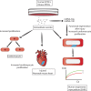
Graphical abstract
Figure 1
Figure 1
EVs isolated from epicardial conditioned media. (A) Brightfield image of mouse epicardial cells in culture with characteristic epithelial morphology. Scale bar denotes 100 µm. (B) Representative size characterization of EVs by nanoparticle tracking. This example shows a particle mode of 93 nm size. (C) Transmission electron micrograph of EVs isolated from epicardial conditioned media. Note the cupped-shaped morphology and size (80 nm). Scale bar denotes 50 nm. (D) CD63 immunogold labelling of EVs isolated from epicardial conditioned media. Data in (B) is presented as frequency of EVs in a given size. Scale bar denotes 50 nm.
Figure 2
Figure 2
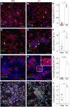
Epicardial EVs promote cell proliferation in mouse neonatal cardiomyocytes. (A, B) Immunostaining of PH3 (green) and α-actinin (red) of cultured mouse neonatal cardiomyocytes treated with vehicle (PBS) (A) or epicardial EVs (EV) (B). Arrowheads in (B) point to PH3 positive cardiomyocytes, co-stained for both markers. (C) Quantification of proportion of cardiomyocytes (α-actinin positive) positive for PH3 in both vehicle (PBS) and EV-treated cultures (EV). (D, E) Immunostaining for BrdU (green) and α-actinin (red) of cultured mouse neonatal cardiomyocytes, treated with vehicle (PBS) (D) or epicardial EVs (EV) (E). (F) Quantification of proportion of cardiomyocytes (α-actinin positive) positive for BrDU in both vehicle (PBS) and EV-treated cultures (EV). (G, H) Immunostaining for Aurora-B (green) to show cytokinesis and α-actinin (red) of cultured mouse neonatal cardiomyocytes, treated with vehicle (PBS) (G) or epicardial exosomes (Exo). (H) Magnification of boxed area and arrowhead in (H) point to a positive cytokinetic furrow between cardiomyocyte cells. Note only immunoreactivity during cytokinesis is considered as positive Aurora-B signal. (I) Quantification of proportion of cardiomyocytes (α-actinin positive) positive for PH3 in both vehicle (PBS) and EV-treated (EV) cultures. (J, K) Immunostaining α-actinin (grey) of cultured mouse neonatal cardiomyocytes from FUCCI line, in which endogenous Venus (green) fluorescence labels cells in S-M and Tomato, (red), cells in G1 phase of the cell cycle; treated with vehicle (PBS) (J) or epicardial EVs (EV) (K). (L) Quantification of proportion of cardiomyocytes (α-actinin positive) positive for Venus-FUCCI in both vehicle (PBS) and EV-treated (EV) cultures. DAPI (blue) labels cell nuclei. Data are presented as mean±SEM. n=4 PH3, Control; n=4 PH3, EVs; n=5 BrdU, Control; n=5 BrdU, EVs; n=3 Aurora-B, Control; n=5 Aurora-B, EVs; n=4 FUCCI, Control; n=4 FUCCI, EVs. *P<0.05, **P<0.01, ***P<0.001, ****P<0.0001. Mann–Whitney test. Scale bars: 50 µm.
Figure 3
Figure 3
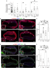
Epicardial EVs increase cardiomyocyte proliferation after injury in P1 and P7 hearts. (A) α-actinin (grey) and FUCCI reporter (green and red) detection in the neonatal heart. DAPI (blue) labels cell nuclei. Left panel shows merge of all the channels and includes orthogonal views to highlight detection of cardiomyocyte nuclei positive for the FUCCI reporters. Middle and right panels are separate channels from the magnified boxed area in the left panel. Empty arrowhead points to a Venus (green) positive cardiomyocyte. Filled arrowhead points to a tomato (red) positive cardiomyocyte. Filled arrow points to a cardiomyocyte displaying both FUCCI markers. Asterisk points to a cardiomyocyte that is not labelled for any of the FUCCI reporters. (BE) FUCCI cell cycle reporter (red and green) and DAPI to label cell nuclei are shown in P8 heart sections collected 7 days after sham or MI procedure followed by epicardial EV or vehicle injection performed at P1. (B) Sham, (C) MI, (D) Sham with administration of EVs, (E) MI with administration of EVs. (F) Quantification of cardiomyocytes’ cell cycle reported by FUCCI expression at P8, 7 days after procedure at P1 (S: sham, MI: myocardial infarction, MI+ EV: myocardial infarction followed by EV injection, S+EV: Sham + EV injection). (GJ) FUCCI cell cycle reporter (red and green) and DAPI to label cell nuclei are shown in P14 heart sections collected 7 days after sham or MI procedure followed by exosome or vehicle injection performed at P7. (G) sham, (H) MI, (I) sham with administration of exosomes, (J) MI with administration of exosomes. (K) Quantification of cardiomyocytes’ cell cycle reported by FUCCI expression at P14, 7 days after procedure at P7 (S, sham; MI, myocardial infarction; MI+ EV, myocardial infarction followed by EV injection; S+EV, Sham + EV injection; LV, left ventricle; LA, left atria; RV, right ventricle; RA, right atria; epi, epicardium; myo, myocardium; end, endocardium). Data are presented as mean SEM. n=3 ShamP1D7; n=4 MI P1D7; n=5 MI+EVsP1D7; n=4, Sham+EVsP1D7; n=3 ShamP7D7; n=5 MI P7D7; n=4 MI+EVsP7D7; n=4, Sham+EVsP7D7. *P<0.05, **P<0.01, ***P<0.001. Mann–Whitney test. Scale bars: 50 µm except magnified panels in (A): 20 µm.
Figure 4
Figure 4
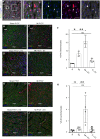
Epithelial and mesenchymal human epicardial cells signal through EV release. (A) Brightfield image of human epicardial cells in culture after expansion from patient RAA biopsy, displaying epithelial morphology. (B) Brightfield image of human epicardial cells in culture after expansion from patient RAA biopsy, displaying mesenchymal spindle morphology after spontaneous activation in vitro. (C) Size characterization of EVs by nanoparticle-tracking graph showing a mode of particle size of 93 nm. (D) TEM image of EVs isolated from epicardial conditioned media. Note the cupped-shaped morphology and size (80 nm). Data in (C) is presented as frequency of EVs in a given size. Scale bars: (A) 100 µm; (D) 100 nm.
Figure 5
Figure 5
Enhanced functional regeneration of EHM after cryoinjury with human epicardial EV treatment. (A) Quantification of maximum contraction forces of EHM after cryoinjury and treatment with human epicardial EVs at day of procedure (d0), 3 days post-procedure (d3), and 7 days post-procedure (d7). (B) α-actinin (red) and PH3 (green) confocal images of EHMs at Day 3 after procedure for the four experimental groups. Arrowheads point to PH3 positive cardiomyocytes. Boxed areas show magnification of representative cells displaying co-staining of PH3 and actinin. Dashed lines surround the cryoinjured EHM arm, displaying cardiomyocyte loss. (C) Graph showing the percentage of cardiomyocytes expressing PH3 in EHMs (C, control; Cry, cryoinjured; E, EVs; Cry+E, cryoinjured treated with EVs). (D) α -SMA (red) and vimentin (green) staining of EHMs 3 days after procedure. Arrowheads point to a-SMA positive fibroblasts (vimentin positive cells). Boxed areas show magnification of representative cells displaying co-staining of vimentin and a-SMA. (E) Quantification of the percentage of vimentin cells co-expressing α -SMA in EHMs (C, control; Cry, cryoinjured, E, EVs; Cry+E, cryoinjured treated with EVs). Data are presented as mean±SEM. n= 4 control d3; n=3 EV-treated d3; n=4 cryoinjured d3; n=4 cryoinjured EV-treated d3; n=4 control d3; n=3 EV-treated d3; n=4 cryoinjured d3; n=4 cryoinjured EV-treated d3. For the maximum force analysis n =44 control n=8 EV d0 n=19 cryo d0 n=4 cryo+EV d0; n=4 EV d3 n=10 cryo d3 n=4 EV+cryo d3; n=4 EV d7 n=12 cryo d7 n=4 EV cryo d7. *P<0.05, for post ANOVA comparisons with control group #P<0.05 for t-test for differences between cryoinjured (Cry) and cryoinjured treated with human epicardial EVs (Cry+E). Scale bars: 200 µm. Dashed lines in (B) and (D) right panels show the loss of cardiomyocytes upon cryoinjury.
Figure 6
Figure 6
miRNA cargo in epicardial EVs is highly conserved in mouse and human; but slight differences are observed among spindle and cobble human epicardial exosomes. (A) Venn diagram summarizing the results from miRNA Sequencing of human (spindle and cobble) and mepic EV. List at the right hand-side shows highly represented miRNA with cardiomyocyte proliferative potential that were taken forward and used in AAV synthesis. miRNAs listed in green are exclusive for human spindle (activated) epicardial exosomes.
Figure 7
Figure 7
miRNAs present in epicardial EVs partially recapitulate the proliferative ability of EV treatment in cardiomyocytes. (AF) Immunostaining of PH3 (green) and α-actinin (red) of cultured mouse neonatal cardiomyocytes, treated with vehicle (A), AAV miR-30a (B), AAV miR-100 (C), AAV miR-27a (D), AAV miR-30e (E). Arrowheads point at proliferating cardiomyocytes, co-stained with a-actinin and PH3. DAPI (blue) labels cell nuclei. (F) Quantification of proliferating cardiomyocytes shown in (AF) represented as percentage of cardiomyocytes positive for PH3 staining after treatment with lentiviral particles. (G) Quantification of maximum contraction forces of EHM after cryoinjury and treatment with AAV miR-100 and AAV miR-30a at 7 days post-procedure. Data are presented as mean SEM. n=3/condition and n>3000 cells/condition for (AF); and n=44 control; n=2 cryoinjured; n=4 AAVmiR-100; n=4 miR30a. *P<0.05, **P <0.01, ***P<0.001, ***P<0.0001. Student’s t-test. Scale bars: 50 µm.

Similar articles

Cited by

References

    1. Go AS, Mozaffarian D, Roger VL, Benjamin EJ, Berry JD, Blaha MJ, Dai S, Ford ES, Fox CS, Franco S, Fullerton HJ, Gillespie C, Hailpern SM, Heit JA, Howard VJ, Huffman MD, Judd SE, Kissela BM, Kittner SJ, Lackland DT, Lichtman JH, Lisabeth LD, Mackey RH, Magid DJ, Marcus GM, Marelli A, Matchar DB, McGuire DK, Mohler ER III, Moy CS, Mussolino ME, Neumar RW, Nichol G, Pandey DK, Paynter NP, Reeves MJ, Sorlie PD, Stein J, Towfighi A, Turan TN, Virani SS, Wong ND, Woo D, Turner MB; American Heart Association Statistics Committee, Stroke Statistics Subcommittee. Heart disease and stroke statistics–2014 update: a report from the American Heart Association. Circulation 2014;129:e28–e292. - PMC - PubMed
    1. Bergmann O, Bhardwaj RD, Bernard S, Zdunek S, Barnabé-Heider F, Walsh S, Zupicich J, Alkass K, Buchholz BA, Druid H, Jovinge S, Frisén J.. Evidence for cardiomyocyte renewal in humans. Science 2009;324:98–102. - PMC - PubMed
    1. Bollini S, Riley PR, Smart N.. Thymosin beta4: multiple functions in protection, repair and regeneration of the mammalian heart. Expert Opin Biol Ther 2015;15:163– 174. - PubMed
    1. Smart N, Bollini S, Dube KN, Vieira JM, Zhou B, Davidson S, Yellon D, Riegler J, Price AN, Lythgoe MF, Pu WT, Riley PR.. De novo cardiomyocytes from within the activated adult heart after injury. Nature 2011;474:640–644. - PMC - PubMed
    1. Simoes FC, Riley PR.. The ontogeny, activation and function of the epicardium during heart development and regeneration. Development 2018;145:dev155994. - PubMed

Publication types

MeSH terms