Multicenter Study
. 2021 Jun 1;6(6):624-632.
doi: 10.1001/jamacardio.2021.0185.

Utility of a Deep-Learning Algorithm to Guide Novices to Acquire Echocardiograms for Limited Diagnostic Use

Affiliations
Multicenter Study

Utility of a Deep-Learning Algorithm to Guide Novices to Acquire Echocardiograms for Limited Diagnostic Use

Akhil Narang et al. JAMA Cardiol. .

Abstract

Importance: Artificial intelligence (AI) has been applied to analysis of medical imaging in recent years, but AI to guide the acquisition of ultrasonography images is a novel area of investigation. A novel deep-learning (DL) algorithm, trained on more than 5 million examples of the outcome of ultrasonographic probe movement on image quality, can provide real-time prescriptive guidance for novice operators to obtain limited diagnostic transthoracic echocardiographic images.

Objective: To test whether novice users could obtain 10-view transthoracic echocardiographic studies of diagnostic quality using this DL-based software.

Design, setting, and participants: This prospective, multicenter diagnostic study was conducted in 2 academic hospitals. A cohort of 8 nurses who had not previously conducted echocardiograms was recruited and trained with AI. Each nurse scanned 30 patients aged at least 18 years who were scheduled to undergo a clinically indicated echocardiogram at Northwestern Memorial Hospital or Minneapolis Heart Institute between March and May 2019. These scans were compared with those of sonographers using the same echocardiographic hardware but without AI guidance.

Interventions: Each patient underwent paired limited echocardiograms: one from a nurse without prior echocardiography experience using the DL algorithm and the other from a sonographer without the DL algorithm. Five level 3-trained echocardiographers independently and blindly evaluated each acquisition.

Main outcomes and measures: Four primary end points were sequentially assessed: qualitative judgement about left ventricular size and function, right ventricular size, and the presence of a pericardial effusion. Secondary end points included 6 other clinical parameters and comparison of scans by nurses vs sonographers.

Results: A total of 240 patients (mean [SD] age, 61 [16] years old; 139 men [57.9%]; 79 [32.9%] with body mass indexes >30) completed the study. Eight nurses each scanned 30 patients using the DL algorithm, producing studies judged to be of diagnostic quality for left ventricular size, function, and pericardial effusion in 237 of 240 cases (98.8%) and right ventricular size in 222 of 240 cases (92.5%). For the secondary end points, nurse and sonographer scans were not significantly different for most parameters.

Conclusions and relevance: This DL algorithm allows novices without experience in ultrasonography to obtain diagnostic transthoracic echocardiographic studies for evaluation of left ventricular size and function, right ventricular size, and presence of a nontrivial pericardial effusion, expanding the reach of echocardiography to clinical settings in which immediate interrogation of anatomy and cardiac function is needed and settings with limited resources.


Conflict of interest statement

Conflict of Interest Disclosures: Dr Hong, Ms Y. Thomas, Mr Surette, Dr Cadieu, Mr Chaudhry, and Dr Martin are employees of Caption Health Inc. Drs Rubenson, Goldstein, Little, Lang, Weissman, and J. Thomas were paid consultants for Caption Health Inc for this study. Dr J. Thomas reports spouse employment at Caption Health Inc. Dr Narang previously received honoraria from Caption Health Inc for an unrelated study. Dr Hong also reported personal fees from Caption Health Inc during the conduct of the study and outside the submitted work, patents US20180153505A1 and US20200245970A1 pending, and patents US10631791B2, US10470677B2, and US10726548B2 issued. Mr Surette reported patent US10726548 issued with Bay Labs Inc, the former name of Caption Health Inc. Dr Cadieu reported personal fees from Caption Health Inc, including stock ownership, during the conduct of the study and outside the submitted work; in addition, Dr Cadieu had patents US20180153505A1 and US20200245970A1 pending and patents US10631791B2, US10470677B2, and US10726548B2 issued. Dr Chaudhry reported patent US20200245970A1 pending. Dr Martin reported stock ownership in Caption Health Inc outside the submitted work. Dr McCarthy reported personal fees and royalties from Edwards Lifesciences, other support from Abbott Vascular as a co–principal investigator of the Repair MR Trial, and personal fees (speaker fees or honoraria) from Medtronic and Atricure outside the submitted work. Dr Rubenson reported personal fees from Caption Health during the conduct of the study. Dr Little reported personal fees from Caption Health outside the submitted work. Dr Lang reported personal fees from Caption Health during the conduct of the study and outside the submitted work. Dr Weissman reported grants from Baylabs during the conduct of the study. Dr J. Thomas also reported personal fees from Caption Health during the conduct of the study and grants from GE and Abbott Vascular and personal fees from Edwards and Shire outside the submitted work. No other disclosures were reported.

Figures

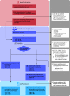
Figure 1.
Figure 1.. Study Design
MHI indicates the Minneapolis Heart Institute; mITT, modified intention to treat; NM, Northwestern Memorial.
Figure 2.
Figure 2.. Representative Still Images of 4 of the 10 Standard Transthoracic Echocardiographic Views Acquired by a Nurse Using the Deep-Learning Algorithm That Were Judged to Be of Diagnostic Quality
All 10 images are in eFigure 3 in the Supplement.

References

    1. Knackstedt C, Bekkers SC, Schummers G, et al. . Fully automated versus standard tracking of left ventricular ejection fraction and longitudinal strain: the FAST-EFS multicenter study. J Am Coll Cardiol. 2015;66(13):1456-1466. doi:10.1016/j.jacc.2015.07.052 - DOI - PubMed
    1. Kusunose K, Abe T, Haga A, et al. . A deep learning approach for assessment of regional wall motion abnormality from echocardiographic images. JACC Cardiovasc Imaging. 2020;13(2 pt 1):374-381. doi:10.1016/j.jcmg.2019.02.024 - DOI - PubMed
    1. Kusunose K, Haga A, Abe T, Sata M. Utilization of artificial intelligence in echocardiography. Circ J. 2019;83(8):1623-1629. doi:10.1253/circj.CJ-19-0420 - DOI - PubMed
    1. Madani A, Arnaout R, Mofrad M, Arnaout R. Fast and accurate view classification of echocardiograms using deep learning. NPJ Digit Med. 2018;1:1. doi:10.1038/s41746-017-0013-1 - DOI - PMC - PubMed
    1. Medvedofsky D, Mor-Avi V, Amzulescu M, et al. . Three-dimensional echocardiographic quantification of the left-heart chambers using an automated adaptive analytics algorithm: multicentre validation study. Eur Heart J Cardiovasc Imaging. 2018;19(1):47-58. doi:10.1093/ehjci/jew328 - DOI - PubMed

Publication types