The Flash-lag Effect in Amblyopia
- PMID: 33599734
- PMCID: PMC7900860
- DOI: 10.1167/iovs.62.2.23
The Flash-lag Effect in Amblyopia
Abstract
Purpose: Amblyopes suffer a defect in temporal processing, presumably because of a neural delay in their visual processing. By measuring flash-lag effect (FLE), we investigate whether the amblyopic visual system could compensate for the intrinsic neural delay due to visual information transmissions from the retina to the cortex.
Methods: Eleven adults with amblyopia and 11 controls with normal vision participated in this study. We assessed the monocular FLE magnitude for each subject by using a typical FLE paradigm: a bar moved horizontally, while a flashed bar briefly appeared above or below it. Three luminance contrasts of the flashed bar were tested: 0.2, 0.6, and 1.
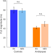
Results: All participants, controls and those with amblyopia, showed a typical FLE. However, the FLE magnitude of participants with amblyopia was significantly shorter than that of the control participants, for both their amblyopic eye (AE) and fellow eye (FE). A nonsignificant difference was found in FLE magnitude between the AE and the FE.
Conclusions: We demonstrate a reduced FLE both in the AE as well as the FE of patients with amblyopia, suggesting a global visual processing deficit. We suggest it may be attributed to a more limited spatiotemporal extent of facilitatory anticipatory activity within the amblyopic primary visual cortex.
Conflict of interest statement
Disclosure:
Figures


References
-
- Bedell HE, Flom MC, Barbeito R.. Spatial aberrations and acuity in strabismus and amblyopia. Invest Ophthalmol Vis Sci . 1985; 26(7): 909–916. - PubMed
-
- Hess RF, Howell ER.. The threshold contrast sensitivity function in strabismic amblyopia: evidence for a two type classification. Vision Res . 1977; 17(9): 1049–1055. - PubMed
-
- Hess RF, Campbell FW, Greenhalgh T.. On the nature of the neural abnormality in human amblyopia; neural aberrations and neural sensitivity loss. Pflugers Arch . 1978; 377(3): 201–207. - PubMed
-
- Hess RF, Holliday IE.. The spatial localization deficit in amblyopia. Vision Res . 1992; 32(7): 1319–1339. - PubMed
-
- Spang K, Fahle M.. Impaired temporal, not just spatial, resolution in amblyopia. Invest Ophthalmol Vis Sci . 2009; 50(11): 5207–5212. - PubMed
Publication types
MeSH terms
Grants and funding
LinkOut - more resources
Full Text Sources
Other Literature Sources
Medical

