Review
. 2021 Nov;51(12):2270-2283.
doi: 10.1007/s00247-021-04974-4. Epub 2021 Feb 18.

Contrast-enhanced ultrasound of the pediatric brain

Affiliations
Review

Contrast-enhanced ultrasound of the pediatric brain

Misun Hwang et al. Pediatr Radiol. 2021 Nov.

Abstract

Brain contrast-enhanced ultrasound (CEUS) is an emerging application that can complement gray-scale US and yield additional insights into cerebral flow dynamics. CEUS uses intravenous injection of ultrasound contrast agents (UCAs) to highlight tissue perfusion and thus more clearly delineate cerebral pathologies including stroke, hypoxic-ischemic injury and focal lesions such as tumors and vascular malformations. It can be applied not only in infants with open fontanelles but also in older children and adults via a transtemporal window or surgically created acoustic window. Advancements in CEUS technology and post-processing methods for quantitative analysis of UCA kinetics further elucidate cerebral microcirculation. In this review article we discuss the CEUS examination protocol for brain imaging in children, current clinical applications and future directions for research and clinical uses of brain CEUS.

Keywords: Brain; Children; Contrast-enhanced ultrasound; Epilepsy; Hypoxic–ischemic injury; Neurovascular disease; Stroke; Tumor; Ultrasound; Ultrasound contrast agents.


Conflict of interest statement

Conflicts of interest None

Figures

Fig. 1
Fig. 1
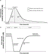
Contrast-enhanced ultrasound (CEUS) quantification methods: bolus-based time-intensity curve and infusion-based destruction-replenishment method. a Representative plot of a time-intensity curve generated from bolus administration of ultrasound contrast agent (UCA). The contrast agent intensity is represented by the solid line, with the intensity amplitude in arbitrary units (au) or decibels (dB) on the y-axis and time on the x-axis. Also shown are perfusion kinetics metrics of arrival time, time to peak, wash-in slope, wash-in area under the curve, washout area under the curve, washout slope and peak intensity that can be obtained from the time-intensity curve. b Representative plot of the UCA intensity changes over time using an infusion-based destruction–replenishment method. After infusion is initiated and using low mechanical index (MI) imaging, the UCA intensity gradually increases until it reaches a steady-state enhancement (solid line). A high mechanical index pulse is then delivered, causing microbubble destruction within the imaging area of interest. The mechanical index is returned to a low setting to allow preservation of microbubbles washing into the field of view. The initial slope on the replenishment cycle can be used as an indicator of flow-rate or flux rate
Fig. 2
Fig. 2
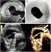
Contrast-enhanced ultrasound (CEUS) in normal brain and in hypoxic–ischemic injury. a Brain CEUS in a normal 1-month-old boy in the coronal plane demonstrates more avid perfusion to the central gray nuclei (CN) as compared to white matter (WM) during the wash-in phase. b Brain CEUS in a 14-day-old boy with known hypoxic–ischemic injury to the white matter. Image obtained in the coronal plane during the peak enhancement phase shows hyperenhancement of the internal capsule (IC) and corpus callosum (CC) during the reperfusion state. The degree of enhancement is like that of the central gray nuclei (CN), resulting in loss of differential perfusion between these structures. Central gray nuclei should be the most avidly enhancing structure at peak enhancement in infants. c Brain CEUS in a 10-month-old girl with known multiple hypoxic–ischemic insults to the brain. Image obtained in the coronal plane during wash-in phase shows mild ventriculomegaly (V) and avid perfusion of the cortical ribbon (dashed line), corpus callosum (CC) and central gray nuclei (CN). This finding predates the extensive laminar necrosis on follow-up MRI (not shown here)
Fig. 3
Fig. 3
Maximum-intensity projection (MIP) images of cerebral vasculature. a–d Coronal MIP images of the cerebral vasculature in a 31-day-old girl with transposition ofthe great arteries during the arterial phase depict normal progressive arrival of the US contrast agent from 0 s (a) to 30 s (b), 36 s (c) and 54 s (d) post injection. Numbers on the upper aspect of each image represent the time, in seconds, following the administration of the US contrast agent
Fig. 4
Fig. 4
Visualization of dynamic intracerebral flow characteristics. a Exemplary transfontanelle coronal contrast-enhanced ultrasound (CEUS) images in a 31-day-old girl with transposition of the great arteries taken before surgical intervention. Regions of interest (ROI) were set for both hemispheres (green), the right hemisphere (purple), the left hemisphere (white), and the superior sagittal sinus (yellow). b, c The corresponding color-coded maps in the coronal plane show the washin–washout area-under-the-curve signal (b) and the time to peak (c). Mild asymmetry in cerebral perfusion is evident. d Time-intensity curve generated from the ROIs. X-axis shows time in seconds and y-axis shows signal intensity in arbitrary units (a.u.). Graphs are shown for the right hemisphere (purple), left hemisphere (gray) and the superior sagittal sinus (yellow). Both hemispheres show similar flow patterns with similar quantitative parameters (time to peak is shown). In contrast, venous flow in the superior sagittal sinus shows later enhancement (increased time to peak), as expected
Fig. 5
Fig. 5
Fusion imaging for virtual navigation between the preoperative MRI and real-time contrast-enhanced ultrasound (CEUS), which was performed through a temporal craniotomy in a 15-year-old boy with dysembrioplastic neuroepithelial tumor (DNET). a Axial intraoperative CEUS image (near-field: right temporal lobe). b Co-planar axial fluid-attenuated inversion recovery MRI. The DNET in the medial right temporal lobe shows high intensity (arrows in a). The two imaging modalities are linked and the preoperative MRI follows the real-time CEUS as the US probe is tracked in the three-dimensional space. On CEUS the tumor shows a similar vascularization (oval) compared to the surrounding parenchyma (rectangle). Other anatomical structures visible on CEUS: the mesencephalon (asterisk), the cavernous sinus (arrowhead) and the basilar artery (solid arrow), and the posterior cerebral arteries (open arrow)
Fig. 6
Fig. 6
Contrast-enhanced ultrasound (CEUS) evaluation of a vein of Galen malformation post endovascular coiling in a 2-month-old boy. a Sagittal T2-weighted MRI brain sequence shows dark flow voids in the large arteriovenous malformation. b Fluoroscopic lateral image of the brain post endovascular coiling of the malformation. c Post-coiling sagittal CEUS with dual display of the gray-scale (left) and contrast-enhanced (right) modes obtained using the anterior fontanelle as the acoustic window to assess for residual flow in the coiled arteriovenous malformation. There is small-volume residual flow within the coiled malformation, with mildly prominent parasagittal vessels (arrows)

References

    1. Hwang M (2018) Introduction to contrast-enhanced ultrasound of the brain in neonates and infants: current understanding and future potential. Pediatr Radiol 49:254–262 - PMC - PubMed
    1. Vinke EJ, Kortenbout AJ, Eyding J et al. (2017) Potential of contrast-enhanced ultrasound as a bedside monitoring technique in cerebral perfusion: a systematic review. Ultrasound Med Biol 43:2751–2757 - PubMed
    1. Archer LN, Levene MI, Evans DH(1986) Cerebral artery Doppler ultrasonography for prediction of outcome after perinatal asphyxia. Lancet 2:1116–1118 - PubMed
    1. Hwang M, Sridharan A, Darge K et al. (2019) Novel quantitative contrast-enhanced ultrasound detection of hypoxic ischemic injury in neonates and infants: pilot study 1. J Ultrasound Med 38: 2025–2038 - PubMed
    1. Prada F, Bene MD, Fornaro R et al. (2016) Identification of residual tumor with intraoperative contrast-enhanced ultrasound during glioblastoma resection. Neurosurg Focus 40:E7 - PubMed

LinkOut - more resources