

The effect of pelvic pathology on uterine vein diameters
- PMID: 33599877
- PMCID: PMC7892655
- DOI: 10.1186/s13089-021-00212-y
The effect of pelvic pathology on uterine vein diameters
Abstract
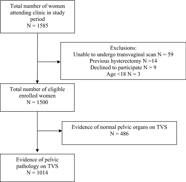
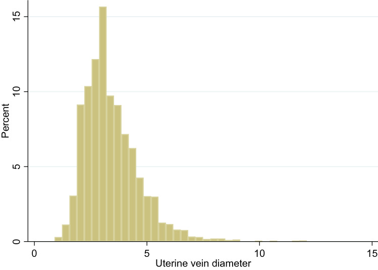
Background: Transvaginal ultrasound (TVS) is a sensitive tool for detecting various conditions that contribute to pelvic pain. TVS can be also used to assess blood flow and measure the size of pelvic veins. Pelvic venous congestion (PVC) is characterised by enlargement of the pelvic veins and has been recognised as a cause of chronic pelvic pain. The reference ranges for uterine venous diameter in women with normal pelvic organs have been established, but there is no information regarding the potential effect of pelvic pathology on the uterine venous diameters. The aim of this study was to examine the size of uterine venous plexus in women with evidence of pelvic abnormalities on TVS and to determine whether the reference ranges need to be adjusted in the presence of pelvic pathology. A prospective, observational study was conducted in our gynaecological outpatient clinic. Morphological characteristics of all pelvic abnormalities detected on TVS and their sizes were recorded. The uterine veins were identified and their diameters were measured in all cases. The primary outcome measure was the uterine venous diameter. Regression analyses were performed to determine factors affecting the uterine venous size in women with pelvic pathology.
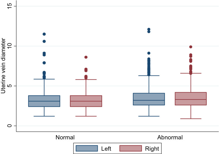
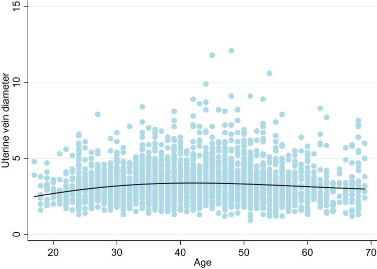
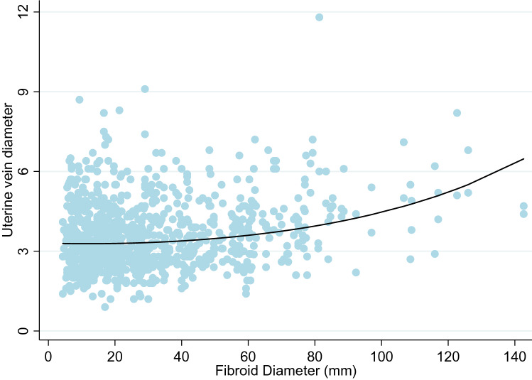
Results: A total of 1500 women were included into the study, 1014 (67%) of whom were diagnosed with pelvic abnormalities. Women with pelvic pathology had significantly larger uterine venous diameters than women with normal pelvic organs (p < 0.01). Multivariable analysis showed that pre-menopausal status, high parity, presence of fibroids (p < 0.001) and Black ethnicity were all associated with significantly larger uterine vein diameters. Based on these findings modified reference ranges for uterine venous diameters have been designed which could be used for the diagnosis of PVC in women with uterine fibroids.
Conclusions: Our findings show that of all pelvic pathology detected on TVS, only fibroids are significantly associated with uterine venous enlargement. Factors known to be associated with enlarged veins in women with normal pelvic organs, namely parity and menopausal status, also apply in patients with pelvic pathology. Future studies of uterine venous circulation should take into account the presence and size of uterine fibroids when assessing women for the signs of PVC.
Keywords: Adenomyosis; Fibroids; Imaging; Pathology; Pelvic venous congestion; Transvaginal ultrasound; Uterine veins.
Conflict of interest statement
The authors declare that they have no competing interests.
Figures
References
-
- Royal College of Obstetricians and Gynaecologists (2012). The Initial Management of Chronic Pelvic Pain (No. 41). Green-top guidelines. London: Royal College of Obstetricians and Gynaecologists
LinkOut - more resources
Full Text Sources
Other Literature Sources