Review
. 2021 Jun;36(5):815-827.
doi: 10.1007/s11011-021-00689-5. Epub 2021 Feb 18.

Cellular and molecular pathophysiology in the progression of Parkinson's disease

Affiliations
Review

Cellular and molecular pathophysiology in the progression of Parkinson's disease

Vandana Zaman et al. Metab Brain Dis. 2021 Jun.

Abstract

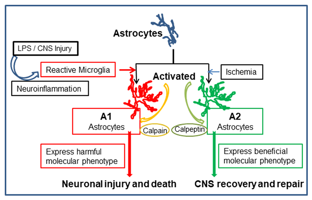
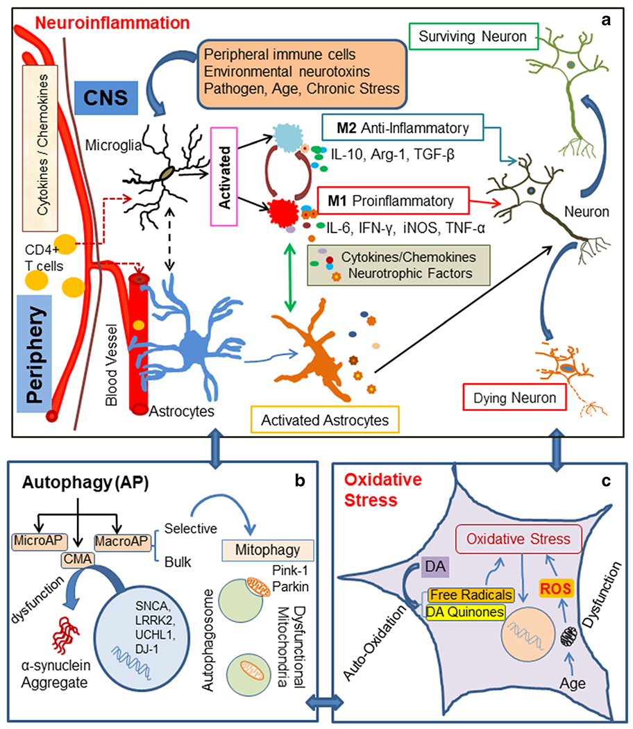
Parkinson's disease (PD) is a neurodegenerative disorder etiologically linked to the loss of substantia nigra (SN) dopaminergic neurons in the mid-brain. The etiopathology of sporadic PD is still unclear; however, the interaction of extrinsic and intrinsic factors may play a critical role in the onset and progression of the disease. Studies in animal models and human post-mortem tissue have identified distinct cellular and molecular changes in the diseased brain, suggesting complex interactions between different glial cell types and various molecular pathways. Small changes in the expression of specific genes in a single pathway or cell type possibly influence others at the cellular and system levels. These molecular and cellular signatures like neuroinflammation, oxidative stress, and autophagy have been observed in PD patients' brain tissue. While the etiopathology of PD is still poorly understood, the interplay between glial cells and molecular events may play a crucial role in disease onset and progression.

Keywords: Autophagy; Neuroinflammation; Neuron; Oxidative damage; Parkinson’s disease; Substantia nigra.


Conflict of interest statement

Conflicts of interest/Competing interests The authors have no financial conflicts of interest.

Figures

Fig. 1
Fig. 1
Schematic representation of astrocyte activation and differentiation following CNS injury by different types of stimuli. Microglia (triggered by different stimuli) contribute to activation of astrocytes. Depending on the micro-environment, astrocytic phenotypic differentiation may result in A1 astrocytes, which contribute to inflammation and neuronal death. However, A2 astrocytes can promote neuronal survival and recovery of function. Calpain activity may facilitate A1 astrocyte differentiation and resulting neuronal injury. However, calpain inhibition by calpeptin may contribute to neuronal survival, CNS recovery and repair through A2 astrocytic activity
Fig. 2
Fig. 2
The diagram depicts several pathways involved in PD pathophysiology, a Neuroinflammation – Microglia are resident immune cells activated by various stimuli, e.g., environmental neurotoxins, pathogens, peripheral inflammation (CD4 + T cells), age, and chronic stress. These stimuli may promote divergent microglial phenotypes such as M1, a proinflammatory phenotype, which can be toxic to neurons. M1-type microglia secrete proinflammatory cytokines such as IL-1β, IL-6, IFN-γ, TNF-α, complement proteins, inducible nitric oxide synthase (iNOS), and reactive oxygen species (ROS). M2, an antiinflammatory phenotype microglia, is believed to be neuroprotective. They secrete cytokines and growth factors such as IL-10, TGF-β, brain-derived neurotrophic factor (BDNF), and arginase-1 (Arg-1). Crosstalk between microglia and astrocytes may promote a time dependent secretion of cytokines/growth factors, contributing to PD pathological conditions, b Autophagy - Neuroinflammation influences all three types of autophagy: (i) macroautophagy (MacroAP), (ii) microautophagy (MicroAP), and (iii) chaperon mediated autophagy (CMA). Genetic mutations may contribute to dysfunction of CMA, leading to α-synuclein aggregation. Mitophagy, a selective autophagy process, can clear dysfunctional mitochondria. Pink1 and Parkin maintain mitochondrial cytoarchitecture and function; they also regulate mitophagy. c Oxidative stress - Autooxidation of dopamine (DA) in dopaminergic neurons generates free radicals and DA quinone. These free radicals lead to oxidative stress. Associated with aging and specific toxins, mitochondrial dysfunction causes the generation of ROS, which ultimately results in oxidative cellular damage

References

    1. Abeliovich A, Gitler AD (2016) Defects in trafficking bridge Parkinson’s disease pathology and genetics. Nature 539:207–216 - PubMed
    1. Ajami B, Bennett JL, Krieger C, Tetzlaff W, Rossi FM (2007) Local self-renewal can sustain CNS microglia maintenance and function throughout adult life. Nat Neurosci 10:1538–1543 - PubMed
    1. Allen NJ, Eroglu C (2017) Cell Biology of Astrocyte-Synapse Interactions. Neuron 96:697–708 - PMC - PubMed
    1. Alliot F, Godin I, Pessac B (1999) Microglia derive from progenitors, originating from the yolk sac, and which proliferate in the brain. Brain Res Dev Brain Res 117:145–152 - PubMed
    1. Alvarez JI, Katayama T, Prat A (2013) Glial influence on the blood brain barrier. Glia 61:1939–1958 - PMC - PubMed

Publication types

MeSH terms

LinkOut - more resources