. 2022 Jun;13(6):446-453.
doi: 10.1007/s13238-021-00828-9. Epub 2021 Feb 18.

Structural and molecular basis for foot-and-mouth disease virus neutralization by two potent protective antibodies

Affiliations

Structural and molecular basis for foot-and-mouth disease virus neutralization by two potent protective antibodies

Hu Dong et al. Protein Cell. 2022 Jun.
No abstract available


Figures

Figure 1
Figure 1
Characterizations of anti-FMDV NAbs M8 and M170. (A) BIAcore SPR kinetic profile of mAb M8 (top) and M170 (bottom) against FMDV O. The binding affinity is depicted by KD (equilibrium dissociation constant, KD = Kd/Ka), which was calculated by the BIAcore 3000 analysis software (BIAevaluation version 4.1). (B) Analysis of the simultaneous binding of M8 and M170 to FMDV through a competitive SPR assay. Due to the requirement of acidic condition for the sensor-labeling, M8 or M170, rather than FMDV, was loaded onto the sensor. In the upper panel, the sensor was labeled with M8. FMDV was inject first, followed by the second injection of M8 again to fully occupy all binding sites for M8 on viral surface, then M170 flowed through. In the bottom panel, the sensor was labeled with M170. The SPR procedure of M170 is same to that of M8. Note: signals (during 800–1000 s) decreased upon M8 binding in the bottom panel, which might result from the ability that M8 has the potential to strip FMDV off the M170-labelled sensor due to the higher binding affinity. (C) Neutralization of FMDV O by M8/M170 (top) and the combination of M8 and M170 (bottom) using plaque-reduction neutralization assay. Neut50 values indicate concentration of antibody required to neutralize fifty percent of the viral titer. The Neut50 of M8 and M170 were 0.8 μmol/L and 3.2 μmol/L, respectively. The Data is presented as the mean ± SD of triplicate measurements. (D) Schematic diagram of M8 or M170 treatment in guinea pig model. Groups of guinea pigs (n = 4) were administrated intramuscularly with M8/M170 (2.5 mg/kg) 1 day before (prophylactic) or after (therapeutic) challenge with 100 ID50 of FMDV on the left hind footpad. Guinea pigs injected intramuscularly with PBS before or after challenge were acted as control groups. The day of FMDV infection was marked as day 0. Protection of guinea pigs against FMDV O by passive immunization with M8 or M170 was analyzed in the prophylactic (Fig. 1E, up) and therapeutic (Fig. 1F, up) modes. No lesions on rear feet were considered as full protection. The copies of virus mRNA in the blood sample from guinea pigs of the prophylactic (Fig. 1E, down) and therapeutic (Fig. 1F, down) groups were quantified by the real-time quantitative PCR (RT-qPCR), the limit of detection (LOD) of viral mRNA in blood was labeled. (G) BIAcore SPR kinetics shows the competitive binding of M8/M170 and αvβ6 integrin to FMDV O. For both panels, αvβ6 integrin was immobilized onto the sensor chips. Mixtures of FMDV O (20 nmol/L) with various concentrations of M8 (up) or M170 (down) acted as running phase to flow through the sensor. Binding signals were detected. (H) Amount of virions remaining on the cell surface, as detected by real-time PCR, when exposed to M8 or M170 before or after the virions attach to BHK21 cells. Data is presented as the mean ± SD. Experiments were repeated in triplicate
Figure 2
Figure 2
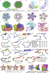
Cryo-EM structures, mechanism of neutralization of FMDV by M8 or M170. (A) Cryo-EM maps of FMDV-M8-complex (left) and FMDV-M170-complex (right). The viral capsids of both complexes are rainbow-colored as the color bar shown below; the M8 and M170 are colored in cyan and magenta, respectively. (B) Electron density maps for the binding interface of M8 and one protomer of the FMDV capsid. The inset shows the density maps and atomic model of VP1 GH loop. Residues with side chains are labeled. Surface representation (left) and epitopes (right) of M8 (C) and M170 (D) on a viral pentamer. The pentamers are shown as surface (left) or cartoon (right) in the signature colors (VP1, blue; VP2, green; VP3, red), while M8 and M170 are colored in the same scheme as in Fig. 2A. The epitopes of M8 (right in 2C) and M170 (right in 2D) are shown as spheres and those from one protomeric unit are circled by black dotted lines. (E) The structural basis of M8 (left) and M170 (right) binding to FMDV. Loops from FMDV involved in interactions with M8/M170 are shown as cartoon, while the remaining parts are shown as surface. M8 and M170 are represented as cartoon. The color scheme is as in Fig. 2A. (F) Detailed interactions between FMDV and M8 (left), as well as FMDV and M170 (right). Residues involved in the binding are shown as sticks and hydrogen bonds are shown as yellow dashed lines. The complementary determining regions of M8 and M170 involved in the interaction with viral capsids are outlined in red and green, respectively. (G) Sequence conservation analysis. The loops from FMDV O that are involved in interactions between FMDV and M8 (VP1 GH loop, VP1 EF loop, VP3 GH loop), as well as FMDV and M170 (VP1 C-terminus, VP3 Knob-βC and VP3 EF loop) are aligned with those of 3 other FMDV serotypes (A, Asia 1 and C) and colored according to sequence conservation as listed in the table below. Residues involved in binding to M8 and M170 are marked by cyan and magenta dots. (H) Structural conservation analysis. The same loops in 2C colored in the signature color scheme are superposed with their counterparts from the apo O (sky blue), A (gray) and C (lime). (I) Clashes between M8 (left)/M170 (right) and αvβ6 receptor. Superimpositions of structures of the FMDV-M8/M170 and FMDV-αvβ6 are shown in pentamer. The capsid pentamer, M8, M170, and receptor subunits αv and β6 are colored in grey, cyan, magenta, bright orange and light pink, respectively. Clashes between M8/M170 and receptor subunits are prominent and marked with star symbols. (J) Clashes between M170 and HS cellular receptor. Color scheme for capsid pentamer and M170 is same as in Fig. 2D. HS receptor is shown as ball and colored in yellow. A protomer together with its corresponding binders (HS and M170) is marked by black lines. The inset shows the clashes between HS and M170

References

    1. Dang M, Wang X, Wang Q, Wang Y, Lin J, Sun Y, Li X, Zhang L, Lou Z, Wang J, et al. Molecular mechanism of SCARB2-mediated attachment and uncoating of EV71. Protein & Cell. 2014;5:692–703. doi: 10.1007/s13238-014-0087-3. - DOI - PMC - PubMed
    1. Harmsen MM, van Solt CB, Fijten HP, van Keulen L, Rosalia RA, Weerdmeester K, Cornelissen AH, De Bruin MG, Eble PL, Dekker A. Passive immunization of guinea pigs with llama single-domain antibody fragments against foot-and-mouth disease. Vet Microbiol. 2007;120:193–206. doi: 10.1016/j.vetmic.2006.10.029. - DOI - PubMed
    1. Harmsen MM, Fijten HPD, Westra DF, Coco-Martin JM. Effect of thiomersal on dissociation of intact (146S) foot-and-mouth disease virions into 12S particles as assessed by novel ELISAs specific for either 146S or 12S particles. Vaccine. 2011;29:2682–2690. doi: 10.1016/j.vaccine.2011.01.069. - DOI - PubMed
    1. Harmsen MM, Seago J, Perez E, Charleston B, Eble PL, Dekker A. Isolation of single-domain antibody fragments that preferentially detect intact (146S) particles of foot-and-mouth disease virus for use in vaccine quality control. Front Immunol. 2017 doi: 10.3389/fimmu.2017.00960. - DOI - PMC - PubMed
    1. Kotecha A, Wang Q, Dong XC, Ilca SL, Ondiviela M, Zihe R, Seago J, Charleston B, Fry EE, Abrescia NGA, Springer TA, Huiskonen JT, Stuart DI (2017) Rules of engagement between αvβ6 integrin and foot-and-mouth disease virus. Nat Commun 8:8 - PMC - PubMed