. 2021 May;31(5):522-539.
doi: 10.1002/hipo.23314. Epub 2021 Feb 18.

Dorsal and ventral mossy cells differ in their axonal projections throughout the dentate gyrus of the mouse hippocampus

Affiliations

Dorsal and ventral mossy cells differ in their axonal projections throughout the dentate gyrus of the mouse hippocampus

Justin J Botterill et al. Hippocampus. 2021 May.

Abstract

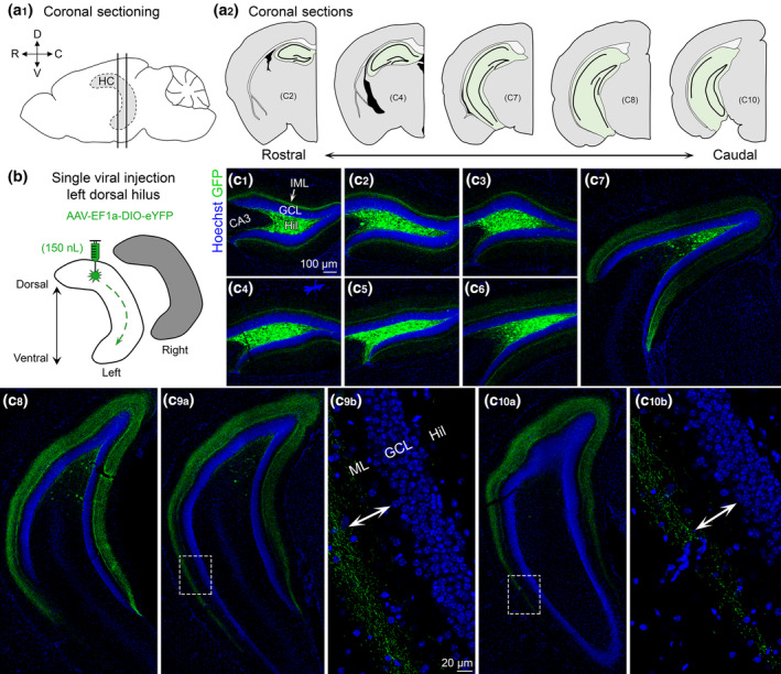
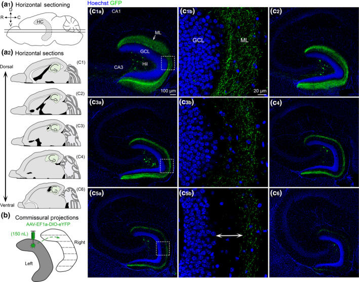
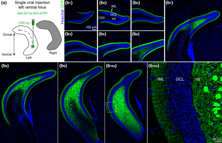
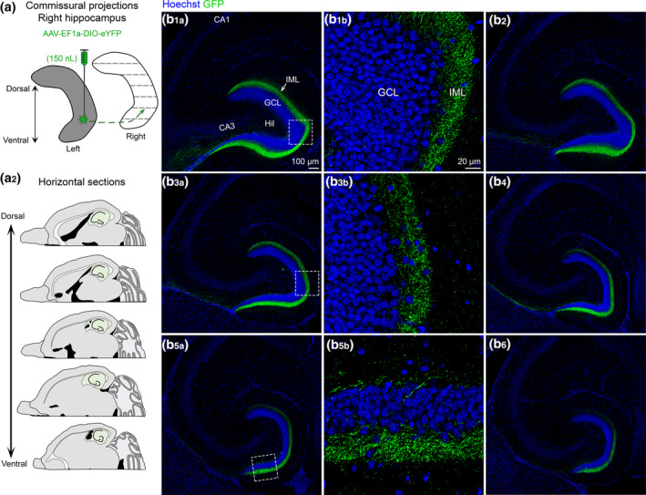
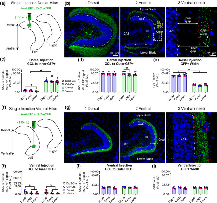
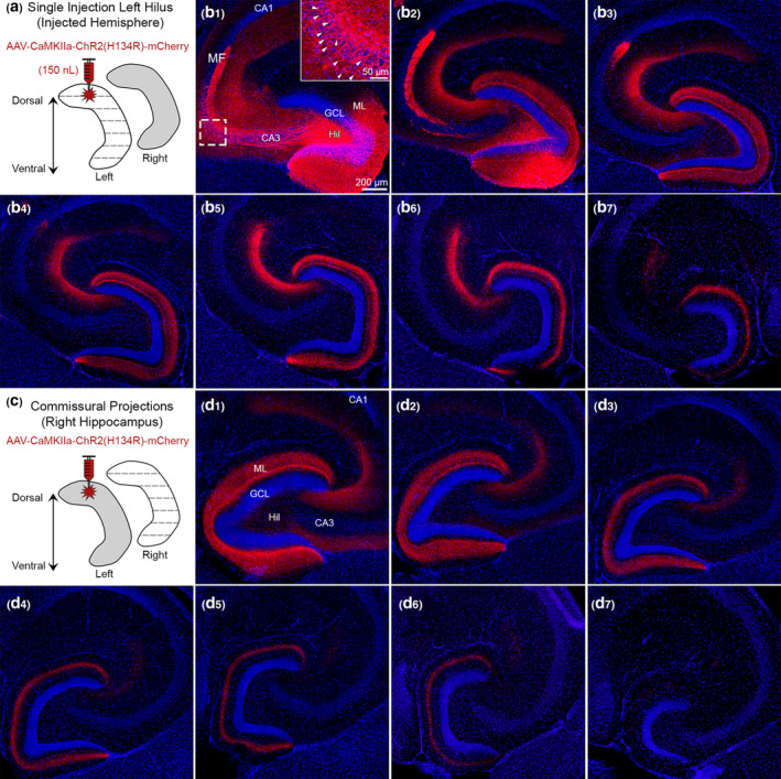
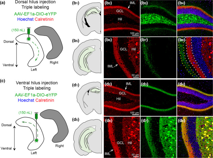
Glutamatergic hilar mossy cells (MCs) have axons that terminate both near and far from their cell body but stay within the DG, making synapses primarily in the molecular layer. The long-range axons are considered the primary projection, and extend throughout the DG ipsilateral to the soma, and project to the contralateral DG. The specificity of MC axons for the inner molecular layer (IML) has been considered to be a key characteristic of the DG. In the present study, we made the surprising finding that dorsal MC axons are an exception to this rule. We used two mouse lines that allow for Cre-dependent viral labeling of MCs and their axons: dopamine receptor D2 (Drd2-Cre) and calcitonin receptor-like receptor (Crlr-Cre). A single viral injection into the dorsal DG to label dorsal MCs resulted in labeling of MC axons in both the IML and middle molecular layer (MML). Interestingly, this broad termination of dorsal MC axons occurred throughout the septotemporal DG. In contrast, long-range axons of ventral MCs terminated in the IML, consistent with the literature. Taken together, these results suggest that dorsal and ventral MCs differ significantly in their axonal projections. Since MC projections in the ML are thought to terminate primarily on GCs, the results suggest a dorsal-ventral difference in MC activation of GCs. The surprising difference in dorsal and ventral MC projections should therefore be considered when evaluating dorsal-ventral differences in DG function.

Keywords: GABA; axon; commissural; hilus; long-range; molecular layer; virus.


Conflict of interest statement

The authors declare that the research was conducted in the absence of any commercial or financial interests that could be construed as a potential conflict of interest.

Figures

FIGURE 1
FIGURE 1
Viral expression of dorsal MCs and axons across the septotemporal axis of the DG. (a1) Side view of the brain showing the hippocampus (HC; grey with dashed border). Straight vertical lines are shown to depict sectioning in the coronal plane. (D) Dorsal, (V) Ventral, (R) Rostral, and (C) Caudal. (a2) Representative schematic of coronal sections starting from the rostral pole and extending to caudal hippocampus (green). (b) Viral injection schematic. 150 nl of AAV‐EF1a‐DIO‐eYFP was injected into the left dorsal hilus. The long‐range axons of MCs are illustrated schematically with green dashes in the left hippocampus. Contralateral projections (right hippocampus; grey) are addressed in Figure 2. (c) Representative viral expression of MCs and their axons (GFP) across the septotemporal DG in a female Crlr‐Cre+/− mouse. Hoechst counterstain (blue) was used to show the DG cell layer. (c1–c4) In the dorsal hippocampus, GFP+ expression was primarily restricted to the hilus and the inner molecular layer (IML). (c5–c8) In progressively more caudal sections, fewer hilar GFP+ cells were observed, but the width of GFP+ axons in the molecular layer increased. (c9–c10) In the most caudal sections, we evaluated, the GFP+ axons terminated in the middle molecular layer in the ventral locations (insets, arrows). GCL, granule cell layer; HIL, hilus; ML, molecular layer [Color figure can be viewed at wileyonlinelibrary.com]
FIGURE 2
FIGURE 2
Contralateral projections of dorsal MC axons across the septotemporal axis of the DG. (a1) Side view of the brain showing the septotemporal extent of the hippocampus (HC; grey with dashed border). Straight horizontal lines are shown to illustrate the horizontal plane. (D) Dorsal; (V) Ventral; (R) Rostral; (C) Caudal. (a2) Representative schematic of horizontal sections from a dorsal level to a progressively more ventral level (green). (b) To evaluate contralateral projections of dorsal MCs, the left hilus (grey) was injected and the right hippocampus (white) was evaluated in the horizontal plane. (c) Representative viral expression in a female Crlr‐Cre+/− mouse. Viral expression in the contralateral hemisphere is shown from sections that were dorsal and progressively more ventral. (c1–c2) In the relatively dorsal sections, there were GFP+ axons throughout the molecular layer. (c3–c4) “Mid” sections (between the dorsal sections in c1–c2 and the ventral sections in c5–c6) showed GFP+ axons that terminated increasingly further away from the GCL border. (c5–c6) GFP+ axons in ventral sections were primarily in the MML (arrow). GCL, granule cell layer; HIL, hilus; ML, molecular layer [Color figure can be viewed at wileyonlinelibrary.com]
FIGURE 3
FIGURE 3
Viral expression in ventral MCs and their axons across the septotemporal axis of the DG. (a) Viral injection schematic. AAV‐EF1a‐DIO‐eYFP was injected into the left ventral hilus. The long‐range axons of ventral MCs are depicted with the green dashes in the left hippocampus (white). Contralateral projections of ventral MCs (right hippocampus; grey) are addressed in Figure 4. (b) Representative viral expression of ventral MCs and their axons (GFP+) across the septotemporal DG in a female Drd2‐Cre+/− mouse. Hoechst counterstain (blue) was used to show the DG cell layer. (b1–b5) In the dorsal hippocampus, GFP+ expression was primarily restricted to the inner molecular layer (IML). (b6–b8) In sections that were progressively more caudal, GFP+ expression was observed in the hilus and IML in the part of the DG that was more ventral. (b9–b10) In sections that show the ventral DG, GFP+ expression was observed in the hilus and IML. GCL, granule cell layer; HIL, hilus; IML, inner molecular layer [Color figure can be viewed at wileyonlinelibrary.com]
FIGURE 4
FIGURE 4
Contralateral projections of ventral MC axons across the septotemporal axis of the DG. (a1) To evaluate contralateral projections of ventral MCs, the left hilus was injected with AAV‐EF1a‐DIO‐eYFP (grey) and the right hippocampus (white) was evaluated in the horizontal plane. (a2) Representative schematic of horizontal sections from dorsal to more ventral hippocampus (green). (b) A representative example of contralateral GFP+ expression in a female Drd2‐Cre+/− mouse. Sections begin at dorsal levels and progress toward more ventral locations. (b1–b6) The contralateral projections of ventral MCs appear to be primarily restricted to the IML across all sections. HIL, hilus; GCL, granule cell layer; IML, inner molecular layer [Color figure can be viewed at wileyonlinelibrary.com]
FIGURE 5
FIGURE 5
Quantitative analysis of the GFP+ MC axon terminal plexus. (a) Schematic showing that AAV‐EF1a‐DIO‐eYFP was injected into the left dorsal hilus. (b1–b2) A representative example of contralateral GFP+ axon terminals in the (b1) dorsal and (b2) ventral DG of a female Crlr‐Cre+/− mouse. The width of the molecular layer (ML) is depicted by the yellow arrow and the approximate subdivisions of the inner (I), middle (M), and outer (O) are separated by dashes. (b3) A schematic shows the inner, outer, and width measurements for the GFP+ axon plexus. Measurements were made in the center of the upper blade, crest, and center of the lower blade and reported as a % of total ML width. (c) In dorsally injected mice, the GFP+ axon was near the GCL/IML border in dorsal sections, but primarily in the MML of ventral sections. Blue and pink data points represent male and female mice, respectively. (d) The outermost distance of the GFP+ axon was approximately 63–64% of dorsal and ventral sections, indicating the GFP+ axons primarily terminated in the MML. (e) The total width of the GFP+ axon was significantly greater in dorsal than ventral sections. (f) A schematic for additional animals where AAV‐EF1a‐DIO‐eYFP was injected into the left ventral hilus. (g1–g2) A representative example of contralateral GFP+ axons in the relatively (g1) dorsal and (g2) ventral DG of a male Drd2‐Cre+/− mouse. (g3) A schematic showing GFP+ axon measurements. (h) The inner distance did not differ between dorsal and ventral sections, but the parts of the DG differed. (i,j) The outer GFP+ distance and total GFP+ width did not differ between dorsal and ventral sections. Notably, all measurements from ventrally injected mice were below 33%, indicating the axon was restricted to the IML. *p < .05 [Color figure can be viewed at wileyonlinelibrary.com]
FIGURE 6
FIGURE 6
Use of CaMKIIa to probe the specificity of GFP for MCs. (a) Viral injection schematic. A total of 150 nl of AAV‐CaMKIIa‐ChR2(H134R)‐mCherry was injected into the left dorsal hilus to target excitatory neurons. (b1–b2) Near the injection site, viral expression was observed in GCs, MCs, and CA3 pyramidal neurons (inset; white arrowheads). Granule cell mossy fibers (MF) axons were also labeled where they normally project, CA3 stratum lucidum. (b3–b7) Long‐range mCherry+ axons showed a similar pattern of viral expression in the molecular layer as Drd2‐Cre or Crlr‐Cre mice injected in the dorsal DG with a virus to express GFP. (c) Contralateral axons were evaluated in the right hippocampus. (d1–d7) Contralateral mCherry+ axons showed a similar pattern in the molecular layer as Drd2‐Cre and Crlr‐Cre injected with a virus expressing GFP in the dorsal hilus. This figure shows that injection of AAV to express CaMKIIa in the dorsal hilus results in a similar pattern of axon labeling as an injection of AAV to express GFP in MCs. Representative images are from a female Drd2‐Cre−/− mouse. GCL, granule cell layer; HIL, hilus; ML, molecular layer [Color figure can be viewed at wileyonlinelibrary.com]
FIGURE 7
FIGURE 7
Calretinin labels ventral but not dorsal MCs. (a) Viral injection schematic. AAV‐EF1a‐DIO‐eYFP was injected into the left dorsal hilus. (b1–b4) In the dorsal DG, calretinin (red) is primarily in the IML of the DG, whereas viral expression (green) is strong in hilar cells and weak in the IML. (b5–b8) In ventral hippocampus, calretinin expression (red) is in putative hilar MCs and the IML. Long‐range GFP+ axons are observed in the molecular layer adjacent to calretinin immunofluorescence in the IML (dotted borders). Representative images are from a female Crlr‐Cre+/− mouse. (c) Viral injection schematic. AAV‐EF1a‐DIO‐eYFP was injected into the left ventral hilus. (d1–d4) Calretinin (red) and GFP+ long‐range axons are primarily in the IML and appear to colocalize (yellow). (d5–d8) In ventral sections, calretinin (red) and GFP strongly overlap within hilar cell bodies and the IML (yellow). Representative images are from a female Drd2‐Cre+/− mouse. GCL, granule cell layer; HIL, hilus; IML, inner molecular layer [Color figure can be viewed at wileyonlinelibrary.com]

Similar articles

Cited by

References

    1. Amaral, D. G. , Scharfman, H. E. , & Lavenex, P. (2007). The dentate gyrus: Fundamental neuroanatomical organization (dentate gyrus for dummies). Progress in Brain Research, 163, 3–22. - PMC - PubMed
    1. Armstrong, C. , Szabadics, J. , Tamas, G. , & Soltesz, I. (2011). Neurogliaform cells in the molecular layer of the dentate gyrus as feed‐forward gamma‐aminobutyric acidergic modulators of entorhinal‐hippocampal interplay. The Journal of Comparative Neurology, 519, 1476–1491. - PMC - PubMed
    1. Azevedo, E. P. , Pomeranz, L. , Cheng, J. , Schneeberger, M. , Vaughan, R. , Stern, S. A. , … Friedman, J. M. (2019). A role of Drd2 hippocampal neurons in context‐dependent food intake. Neuron, 102, 873–886.e875. - PubMed
    1. Bernstein, H. L. , Lu, Y. L. , Botterill, J. J. , Duffy, A. M. , LaFrancois, J. , & Scharfman, H. (2020). Excitatory effects of dentate gyrus mossy cells and their ability to influence granule cell firing: an optogenetic study in adult mouse hippocampal slices. bioRxiv, 1–105. 10.1101/2020.06.06.137844 - DOI
    1. Bernstein, H. L. , Lu, Y. L. , Botterill, J. J. , & Scharfman, H. E. (2019). Novelty and novel objects increase c‐Fos immunoreactivity in mossy cells in the mouse dentate gyrus. Neural Plasticity, 2019, 1815371. - PMC - PubMed

Publication types

LinkOut - more resources