. 2021 Mar 19;16(3):480-490.
doi: 10.1021/acschembio.0c00832. Epub 2021 Feb 18.

The Structural and Biochemical Basis of Apocarotenoid Processing by β-Carotene Oxygenase-2

Affiliations

The Structural and Biochemical Basis of Apocarotenoid Processing by β-Carotene Oxygenase-2

Sepalika Bandara et al. ACS Chem Biol. .

Abstract

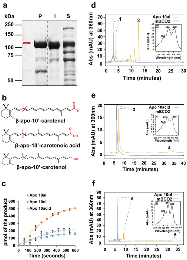
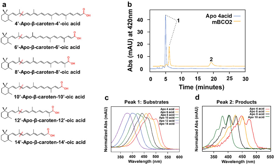
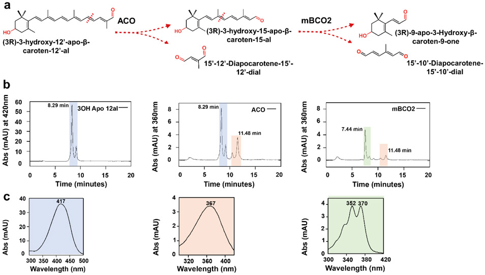
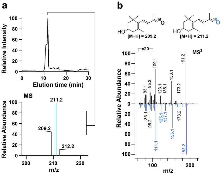
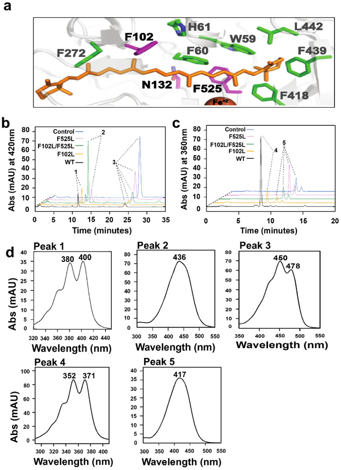
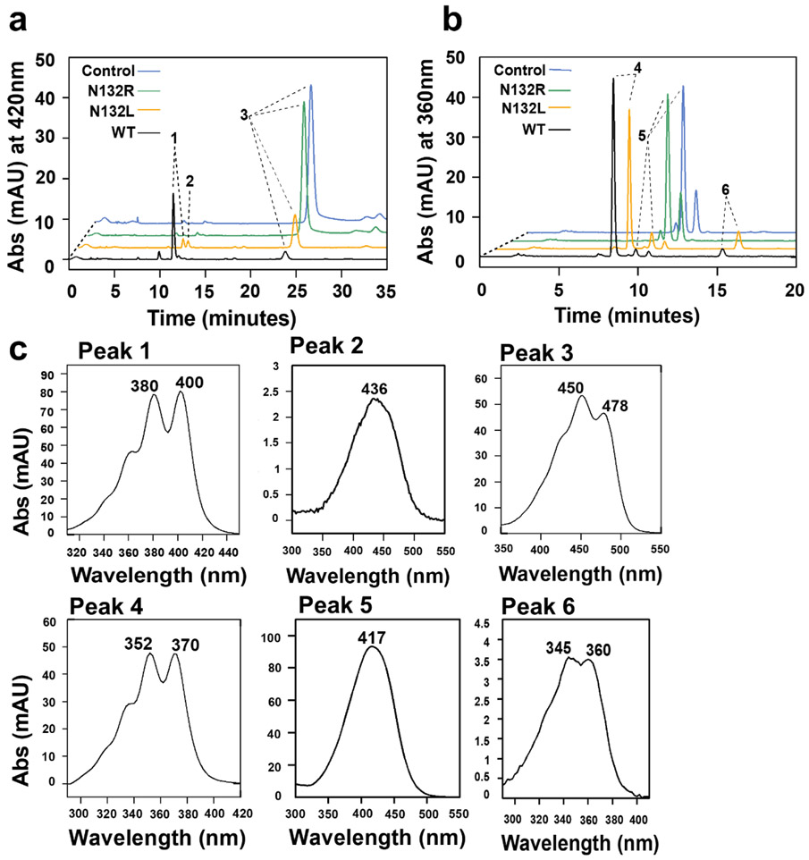
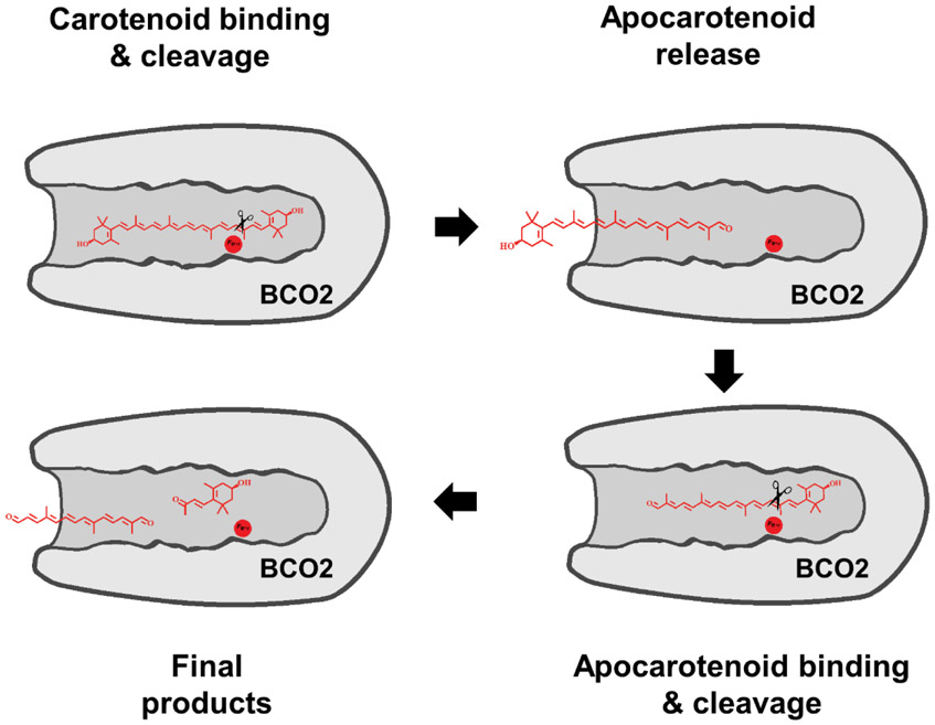
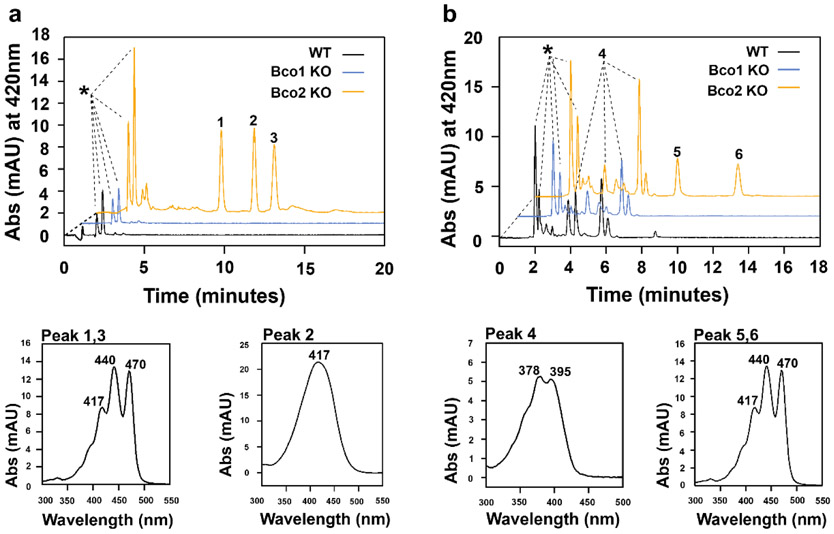
In mammals, carotenoids are converted by two carotenoid cleavage oxygenases into apocarotenoids, including vitamin A. Although knowledge about β-carotene oxygenase-1 (BCO1) and vitamin A metabolism has tremendously increased, the function of β-carotene oxygenase-2 (BCO2) remains less well-defined. We here studied the role of BCO2 in the metabolism of long chain β-apocarotenoids, which recently emerged as putative regulatory molecules in mammalian biology. We showed that recombinant murine BCO2 converted the alcohol, aldehyde, and carboxylic acid of a β-apocarotenoid substrate by oxidative cleavage at position C9,C10 into a β-ionone and a diapocarotenoid product. Chain length variation (C20 to C40) and ionone ring site modifications of the apocarotenoid substrate did not impede catalytic activity or alter the regioselectivity of the double bond cleavage by BCO2. Isotope labeling experiments revealed that the double bond cleavage of an apocarotenoid followed a dioxygenase reaction mechanism. Structural modeling and site directed mutagenesis identified amino acid residues in the substrate tunnel of BCO2 that are critical for apocarotenoid binding and catalytic processing. Mice deficient for BCO2 accumulated apocarotenoids in their livers, indicating that the enzyme engages in apocarotenoid metabolism. Together, our study provides novel structural and functional insights into BCO2 catalysis and establishes the enzyme as a key component of apocarotenoid homeostasis in mice.


Conflict of interest statement

Conflict of interest

The authors declare that they have no conflicts of interest with the contents of this article.

Figures

Figure 1.
Figure 1.
mBCO2 converts β-apocarotenoid substrates with different polar end groups. a) SDS-PAGE of MBP-mBCO2 protein (mBCO2) purification, P indicates protein fractions eluted from the column, I indicates insoluble protein in cell pellet, and S indicates soluble protein in crude extract. Red arrow points to mBCO2 fusion protein. b) Structures of β-apocarotenoids. Functional groups (carboxylic acid, aldehyde, and alcohol) are highlighted in red. c) Enzyme kinetics of mBCO2 (50 μg) with different substrates. The initial velocity was 77.3 pmol x min−1 for β-apo-10’-carotenol, 39.6 pmol x min−1 for β-apo-10’-carotenoic acid, and 21.1 pmol x min−1 for β-apo-10’ carotenal. The experiment was repeated three time and the values are displayed as mean +/− standard deviation. d,e,f) HPLC traces at 360 nm of lipid extracts of enzyme assays with (orange trace) and without (blue trace) mBCO2. The insets show the UV visible spectrum of the respective products. In d) peak 1 is β-apo-10’-carotenal, peak 2 is 10’,10-apocarotene-dialdehyde, in e) peak 3 is β-apo-10’-carotenoic acid, peak 4 is 10-oxo-10,10’-diapocaroten-10’-oic acid, in f) peak 5 is β-apo-10’-carotenol, and peak 6 is 10-Oxo-10,10’-diapocaroten-10’-ol.
Figure 2.
Figure 2.
mBCO2 converts β-apo-carotenoic acids with various chain length. a) Chemical structures of the β-apo-carotenoic acid substrates with different chain length. Expected cleavage sites are indicated with red dashed lines. The carboxylic end groups are highlighted in red. b) Representative HPLC of lipid extracts of reactions with β-apo-4’-carotenoic acid incubated in the presence (orange trace) and absence (blue trace) of mBCO2. Peak 1 is β-apo-4’-carotenoic acid and peak 2 is 10-Oxo-10,4’-diapocarotene-4’-oic acid. c) Representative UV-visible absorption spectra of β-apo-carotenoic acid substrates. d) Representative UV-visible absorption spectra of diapocarotenoid products from enzyme assays with mBCO2.
Figure 3.
Figure 3.
mBCO2 converts 3-hydroxy-retinal after being generated by ACO. a) Reaction scheme for the successive conversion of 3-hydroxy-β-apo-12’-carotenal by ACO to 3-hydroxy-retinal, followed by the conversion of 3-hydroxy-retinal to 3-hydroxy-β-ionone. b) HPLC traces of enzyme assays with ACO and mBCO2. Left panel displays the HPLC trace at 420 nm of the 3-hydroxy-β-apo-12’-carotenal substrate (retention time, 8.29 min); middle panel displays HPLC trace at 360 nm of an enzyme assay with 3-hydroxy-β-apo-12’-carotenal and ACO which results in 3-hydroxy-retinal (retention time, 11.48 min) formation; right panel displays HPLC trace at 360 nm of an enzyme assays in which the 3-hydroxy-retinal produced by ACO was incubated in the presence of mBCO2. Note that this incubation results in the disappearance of the 3-hydroxy-retinal peak. Additionally, 10’,10-apocarotene-dialdehyde (retention time, 7,44 min) is produced by the conversion of the remaining 3-hydroxy β apo-12’-carotenal substrate. c) UV-Visible spectra of peak 1, 3-hydroxy-β-apo-12’-carotenal, peak 2, 3-hydroxy-retinal and peak 3, 12’,10-apocarotene-dialdehyde. The cis isomer of 3-hydroxy-β-apo-12’-carotenal substrate elutes next to the major substrate peak.
Figure 4.
Figure 4.
Murine BCO2 is a dioxygenase. a) LC-MS analysis of 18O2 incorporation into the 3-hydroxy-ionone product of 3-hydroxy- β –apo-12’-carotenal cleavage by mBCO2. b) MS/MS spectra of the two 3-hydroxy-β-ionone isotopologues.
Figure 5.
Figure 5.
BCO2 possesses a bipartite substrate binding cavity which discriminates between carotenoids and apocarotenoids. a) Close up view of the modeled substrate binding cavity of BCO2 with bound β-carotene (orange). The cavity is lined with aromatic amino acid side residues (green) which are conserved between BCO1 and BCO2. Mutated amino acid residue F102, N132 and F525 are highlighted in magenta. b) HPLC traces at 420 nm of enzyme assays of wild type and mutant mBCO2 variants incubated with meso-zeaxanthin. In control experiments, the substrate was incubated with maltose binding protein c) HPLC traces at 360 nm of enzyme assays of wild type and mutant BCO2 variants incubated with 3-hydroxy-β-apo-12’-carotenal. In control experiments, the substrate was incubated with maltose binding protein. d) UV-visible spectra of major products’ peaks in B and C. Peak 1is 10’,10-apocarotene-dialdehyde; peak 2 is 3-β-apo-10’ carotenal, peak 3 is meso-zeaxanthin; peak 4 is 12’,10-apocarotene-dialdehyde; peak 5 is 3-hydroxy-β-apo-12’-carotenal.
Figure 6.
Figure 6.
Asn132 stabilizes π electron bonding in the active center of BCO2. a) HPLC traces at 420 nm of enzyme assays of wild type and mutant mBCO2 variants incubated with meso-zeaxanthin. In control experiments, the substrate was incubated with maltose binding protein b) HPLC traces at 360 nm of enzyme assays of wild type and mutant mBCO2 variants incubated with 3-hydroxy-β-apo-12’-carotenal. In control experiments, the substrate was incubated with maltose binding protein. c) UV-visible spectra of products and substrates. peak 1, 10’,10-apocarotene-dialdehyde; peak 2, beta-apo-10’-carotenal; peak 3, meso-zeaxanthin; peak 4, 12’,10-apocarotene-dialdehyde; peak 5, 3 hydroxy-β-apo-12’-carotenal; peak 6, 10’,10-apocarotene-diol.
Figure 7.
Figure 7.
Cartoon of the different steps of carotenoid cleavage by BCO2. The carotenoid molecule enters the substrate-binding tunnel. Cleaves at the 9,10 double bond results in the removal of one ionone ring site and the formation of an apocarotenoid. The apocarotenoid product leaves the substrate-binding cavity. The apocarotenoid then reenters the substrate-binding cavity in reverse direction followed by cleavage at the 9’,10’ double bond position. The results in the removal of the second ionone ring site and the formation of a diapocarotenoid product.
Figure 8.
Figure 8.
Apocarotenoids accumulate in BCO2-deficient mouse liver. a) HPLC traces at 420 nm of lipid extracts of mice gavaged with 3-hydroxy-β-apo-12’-carotenal. The peaks indicated by asterisks are retinoid peaks (retinyl esters and all-trans-retinol). Lower left panel is UV-visible absorption spectra of unknown dietary compounds (peak 1 and 3) and lower right panel is 3-hydroxy-β-apo-12’-carotenal (peak 2). b) HPLC traces of lipid extracts of mice gavaged with β-apo-10’-carotenal. The peaks indicated by asterisks are retinoid peaks (retinyl esters and all-trans-retinol). Lower left panel is UV-visible absorption spectra of β-apo-10’-carotenol (peak 4) and lower right panel is the two unknown dietary compounds (peaks 5 and 6).

References

    1. Kiser PD, Golczak M, and Palczewski K (2014) Chemistry of the retinoid (visual) cycle, Chemical reviews 114, 194–232. - PMC - PubMed
    1. Kedishvili NY (2013) Enzymology of retinoic acid biosynthesis and degradation, J Lipid Res 54, 1744–1760. - PMC - PubMed
    1. Arshavsky VY, Lamb TD, and Pugh EN Jr. (2002) G proteins and phototransduction, Annu Rev Physiol 64, 153–187. - PubMed
    1. Rhinn M, and Dollé P (2012) Retinoic acid signalling during development, Development (Cambridge, England) 139, 843–858. - PubMed
    1. Hall JA, Grainger JR, Spencer SP, and Belkaid Y (2011) The role of retinoic acid in tolerance and immunity, Immunity 35, 13–22. - PMC - PubMed

Publication types

MeSH terms