Review
. 2021 Apr;27(4):1023-1031.
doi: 10.3201/eid2704.204337. Epub 2021 Feb 18.

Difficulties in Differentiating Coronaviruses from Subcellular Structures in Human Tissues by Electron Microscopy

Review

Difficulties in Differentiating Coronaviruses from Subcellular Structures in Human Tissues by Electron Microscopy

Hannah A Bullock et al. Emerg Infect Dis. 2021 Apr.

Abstract

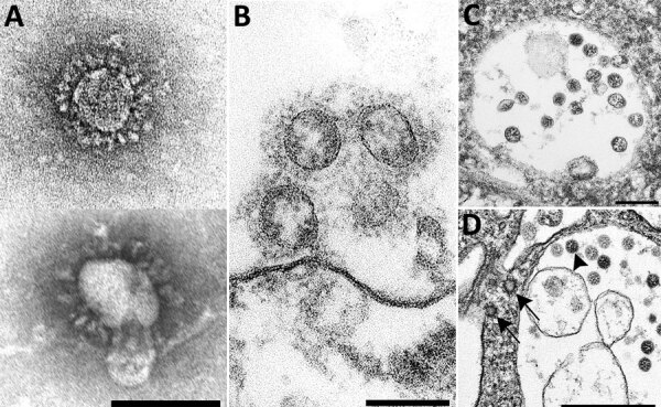
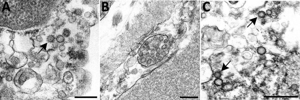
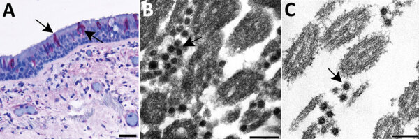
Efforts to combat the coronavirus disease (COVID-19) pandemic caused by severe acute respiratory syndrome coronavirus 2 (SARS-CoV-2) have placed a renewed focus on the use of transmission electron microscopy for identifying coronavirus in tissues. In attempts to attribute pathology of COVID-19 patients directly to tissue damage caused by SARS-CoV-2, investigators have inaccurately reported subcellular structures, including coated vesicles, multivesicular bodies, and vesiculating rough endoplasmic reticulum, as coronavirus particles. We describe morphologic features of coronavirus that distinguish it from subcellular structures, including particle size range (60-140 nm), intracellular particle location within membrane-bound vacuoles, and a nucleocapsid appearing in cross section as dense dots (6-12 nm) within the particles. In addition, although the characteristic spikes of coronaviruses may be visible on the virus surface, especially on extracellular particles, they are less evident in thin sections than in negative stain preparations.

Keywords: COVID-19; SARS; SARS-CoV-2; coronavirus; coronavirus disease; electron microscopy; respiratory infections; severe acute respiratory syndrome coronavirus 2; ultrastructure; viruses; zoonoses.


Figures

Figure 1
Figure 1
Overview of the ultrastructural features of coronavirus morphology as seen by negative stain and thin section. A) Extracellular viral particles ≈100 nm in diameter with prominent peplomers (spikes). Prepared from a cell culture sample by negative stain using heavy metal salt solutions to coat the outside of the virus. Scale bar indicates 100 nm. B) Extracellular viral particles ≈100 nm in diameter with clearly visible spikes. Cross sections through the helical nucleocapsid are visible on the interior of the particle as electron-dense black dots, 6–12 nm in diameter. Prepared by thin section from a formalin-fixed autopsy specimen. Scale bar indicates 100 nm. C) Intracellular viral particles ≈80 nm in diameter held within a membrane-bound vacuole. Cross sections through the helical nucleocapsid are visible inside the particles. Prepared by thin section from a formalin-fixed autopsy specimen. Scale bar indicates 200 nm. D) Intracellular viral particles (arrowhead) within a membrane-bound vacuole and nearby clathrin-coated vesicles (CCV) in the cytoplasm (arrows). CCV spikes directly contact the cell cytosol; viral spikes, barely visible as a faint fuzz, contact the vacuole contents. Cross sections through the helical nucleocapsid are visible inside the viral particles but not within the CCVs. Prepared by thin section from a glutaraldehyde-fixed cell culture sample. Scale bar indicates 500 nm.
Figure 2
Figure 2
Overview of differential ultrastructural features of subcellular structures commonly misidentified as coronaviruses; all were prepared by thin section from formalin-fixed autopsy specimens. A) Clathrin-coated vesicles (CCVs), circular vesicles with a fringe of clathrin protein (arrow), in the cell cytoplasm range in size from 60 nm−100 nm. Differentiation: clathrin surrounding the vesicle may be misinterpreted as viral spikes, however, CCVs are free in the cell cytoplasm, and clathrin is in direct contact with the cytoplasm. Intracellular coronaviruses are found within membrane-bound vacuoles, and spikes, if visible, are in contact with the vacuolar contents. CCVs lack the internal black dots that signify cross sections through the viral nucleocapsid. Scale bar indicates 200 nm. B) Multivesicular body (MVB), a collection of membrane-bound roughly spherical vesicles formed by the inward budding of an endosomal membrane. Differentiation: MVBs may be confused with a vacuolar accumulation of coronavirus particles. Vesicles within multivesicular bodies do not have internal black dots that signify cross sections through the viral nucleocapsid. Scale bar indicates 200 nm. C) Circular cross sections through rough endoplasmic reticulum (RER) (arrows) found free within the cytoplasm. Differentiation: ribosomes along the endoplasmic reticulum may be confused with viral spikes. Ribosomes of vesiculating RER are in direct contact with the cell cytoplasm, unlike coronavirus spikes, which would be in contact with vacuolar contents. Vesiculating RER lacks cross sections through the viral nucleocapsid. Scale bar indicates 1 µm.
Figure 3
Figure 3
Use of immunohistochemistry and electron microscopy to detect severe acute respiratory syndrome coronavirus 2 (SARS-CoV-2) in formalin-fixed paraffin embedded (FFPE) autopsy tissues. A) Immunostaining (arrows) of SARS-CoV-2 in the epithelial cells of the trachea. Scale bar indicates 20 µm. B) Ultrastructural features of extracellular SARS-CoV-2 particles (arrow) in association with ciliated cells of the trachea from paraffin section in panel A, prepared using an FFPE on-slide method. Scale bar indicates 200 nm. C) Thin section of a biopsy punch from the original FFPE block in panel A showing viral particles (arrow) ≈75 nm. Scale bar indicates 200 nm.

References

    1. Yao XH, He ZC, Li TY, Zhang HR, Wang Y, Mou H, et al. Pathological evidence for residual SARS-CoV-2 in pulmonary tissues of a ready-for-discharge patient. Cell Res. 2020;30:541–3. 10.1038/s41422-020-0318-5 - DOI - PMC - PubMed
    1. Pesaresi M, Pirani F, Tagliabracci A, Valsecchi M, Procopio AD, Busardò FP, et al. SARS-CoV-2 identification in lungs, heart and kidney specimens by transmission and scanning electron microscopy. Eur Rev Med Pharmacol Sci. 2020;24:5186–8. - PubMed
    1. Grimes Z, Bryce C, Sordillo EM, Gordon RE, Reidy J, Paniz Mondolfi AE, et al. Fatal pulmonary thromboembolism in SARS-CoV-2 infection. Cardiovasc Pathol. 2020;48:107227. 10.1016/j.carpath.2020.107227 - DOI - PMC - PubMed
    1. Ackermann M, Verleden SE, Kuehnel M, Haverich A, Welte T, Laenger F, et al. Pulmonary vascular endothelialitis, thrombosis, and angiogenesis in Covid-19. N Engl J Med. 2020;383:120–8. 10.1056/NEJMoa2015432 - DOI - PMC - PubMed
    1. Borczuk AC, Salvatore SP, Seshan SV, Patel SS, Bussel JB, Mostyka M, et al. COVID-19 pulmonary pathology: a multi-institutional autopsy cohort from Italy and New York City. Mod Pathol. 2020;33:2156–68. 10.1038/s41379-020-00661-1 - DOI - PMC - PubMed

MeSH terms