Review
. 2021 Jan-Jun:296:100416.
doi: 10.1016/j.jbc.2021.100416. Epub 2021 Feb 16.

Antisense technology: A review

Affiliations
Review

Antisense technology: A review

Stanley T Crooke et al. J Biol Chem. 2021 Jan-Jun.

Abstract

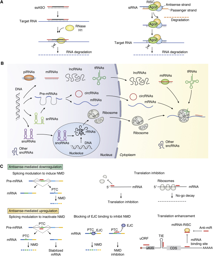
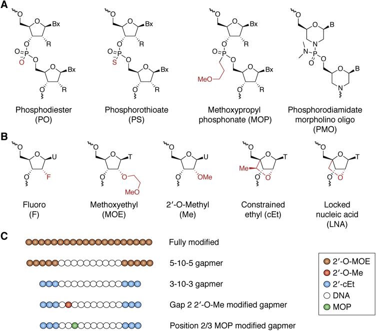
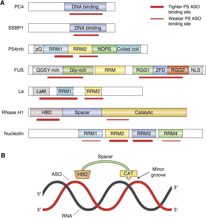
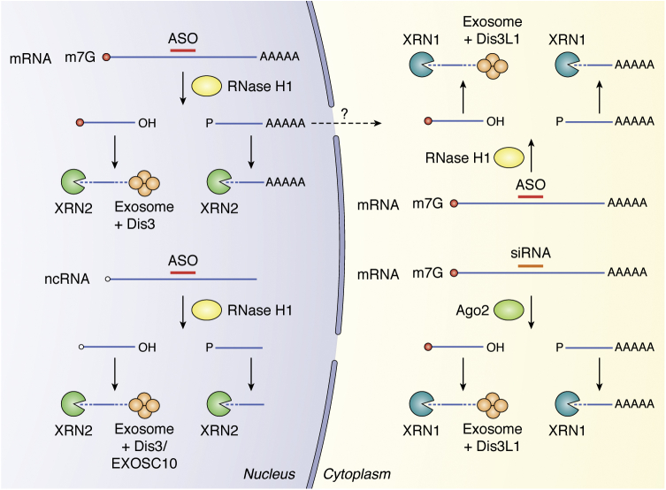
Antisense technology is beginning to deliver on the broad promise of the technology. Ten RNA-targeted drugs including eight single-strand antisense drugs (ASOs) and two double-strand ASOs (siRNAs) have now been approved for commercial use, and the ASOs in phase 2/3 trials are innovative, delivered by multiple routes of administration and focused on both rare and common diseases. In fact, two ASOs are used in cardiovascular outcome studies and several others in very large trials. Interest in the technology continues to grow, and the field has been subject to a significant number of reviews. In this review, we focus on the molecular events that result in the effects observed and use recent clinical results involving several different ASOs to exemplify specific molecular mechanisms and specific issues. We conclude with the prospective on the technology.

Keywords: RNase H1; antisense; clinical results; molecular mechanisms; splicing.


Conflict of interest statement

Conflict of interest The authors declare that they have no conflicts of interest with the contents of this article.

Figures

Figure 1
Figure 1
Antisense technology can modulate gene expression through different mechanisms.A, commonly used antisense mechanisms to degrade target RNAs, including RNase H1-dependent and RISC-dependent mechanisms. B, various types of RNAs. In addition to the three traditional types of RNAs (rRNAs, tRNAs, and mRNAs) that are directly involved in protein synthesis, many types of small noncoding RNAs (<200 nt) have been identified to participate in various biological processes: this includes snRNAs in pre-mRNA splicing, snoRNAs in rRNA processing and modification, miRNAs in translation modulation and mRNA stability, piRNAs in RNA silencing and epigenetics, and other small ncRNAs (e.g., RNase P and MRP RNA) in tRNA and rRNA maturation. Moreover, a large number of long ncRNAs (>200 nt) have also been identified in recent years to play important roles in multiple biological processes, from chromatin remodeling (e.g., Xist, HOTAIR), transcription (e.g., asRNA, eRNA), and RNA processing (e.g., NATS) in the nucleus to translation (e.g., as-Uchl1) and protein modification (e.g., NKILA) in the cytoplasm. In addition, circular RNAs have also been identified that may modulate gene expression by acting as sponges for mRNAs or RNA-binding proteins. C, antisense oligonucleotides can modulate gene expression through additional mechanisms, including downregulation by induced NMD through altering splicing (upper left), inhibit translation initiation by binding to cap region or triggering mRNA no-go decay by binding to coding region of mRNAs (upper right panels). In addition, antisense oligonucleotides can also upregulate gene expression by altering splicing to skip PTC containing exons (lower left panel) or by inhibiting EJC binding to inhibit NMD (lower middle panel) or by enhancing translation through masking translation inhibitory elements, including uORF, TIE, miRNA-binding sites or even miRNAs (lower right panel). The circles of different colors indicate proteins. CDS, coding region sequence; EJC, exon–exon junction complex; NMD, non-sense-mediated decay; PTC, premature termination codon; RISC, RNA-induced silencing complex; TIE, translation inhibit element; uORF, upstream open reading frame. The red bars indicate antisense oligonucleotides or miRNAs.
Figure 2
Figure 2
Oligonucleotide chemical modifications.A, commonly used backbone modifications. MOP, methoxypropyl phosphonate; PMO, phosphorodiamidate morpholino oligomer; PO, phosphodiester; PS, phosphorothioate. B, commonly used 2’ modifications. cEt, constrained ethyl; F, fluoro; LNA, locked nucleic acid; Me, 2’-O-methyl; MOE, methoxyethyl. C, ASO designs. Fully modified ASOs that do not activate RNase H1 are commonly used to modulate splicing and translation. Gapmer ASOs that contain DNA gap and activate RNase H1 are normally modified at both ends of ASOs with different 2’ modifications. To further improve ASO performance, 2’Me or MOP (methoxypropyl phosphonate) modification is introduced into certain positions of the gap region.
Figure 3
Figure 3
Schematic prediction of the domains of model PS-ASO binding proteins.A, protein domains involved in PS-ASO binding. PS-ASO binding domains are induced with red bars. Tighter binding is marked with thicker bars. LaM, La motif; NLS, nuclear localization signal; NOPS, Nona/Paraspeckle domain; pQ, proline and glutamine-rich domain; RGG, arginine/glycine–glycine-rich domain; RRM, RNA recognition motif; ZFD, zinc-finger domain. B, schematic illustration of the relationship of HBD and catalytic domains on a heteroduplex. The HBD and Cat domains act as a caliper to measure approximately one helical turn. This enables the enzyme to bind to any duplex, but discriminate RNA/DNA heteroduplex from RNA/RNA and DNA/DNA duplexes based on slight differences in helical geometry. The catalytic domain measures the width of the minor groove and detects the presence or absence of a 2’OH and phosphate positioned properly. The enzyme is minimally processive, cleaving at most one or two nucleotides 3’ to its initial cleavage site. CAT, catalytic domain; HBD, hybrid binding domain.
Figure 4
Figure 4
RNA cleavage fragments by RNase H1 or siRNA are degraded by 5’-3’ exonucleases and 3’-5’ exonucleases. In the nucleus, the 5’ cleaved fragments are further degraded from 3’ end by exosomes and Dis3 and from 5’ end by XRN2 after decapping. The 3’ fragment is further degraded by 5’-3’ exonuclease XRN2. In the cytoplasm, the 5’ fragment is degraded by XRN1 from 5’ end after decapping and by cytoplasmic exosome and Dis3L1 from 3’ end. The 3’ fragment is degraded from 5’ end by XRN1. Adapted from ref (152).
Figure 5
Figure 5
Many factors affect the activity of PS-ASOs.A, schematic representation of factors that affect PS-ASO activity. Though the level of RNase H1 and the number of PS-ASO binding sites in an RNA positively correlate with ASO activity, other factors, such as RNA structure, RNA-binding protein, or PS-ASO-binding protein, can inhibit PS-ASO activity. In addition, both splicing and translation can affect the activity of PS ASOs. On the other hand, RNA copy number and RNA half-life do not normally affect ASO activity. B, a feedback transcriptional upregulation can cause tolerance effect of gapmer PS ASOs. Some gapmer ASOs targeting coding region of mRNAs can trigger RNase H1 cleavage of the mRNA, leading to enhanced transcription and increased pre-mRNA levels, in an RNase H1, translation, and UPF3A-dependent manner. This in turn can reduce the activity of PS ASOs in decreasing the mRNA levels. Adapted from ref (157).
Figure 6
Figure 6
Mechanisms of PS ASO cellular uptake and release.A, cell surface PS ASO adsorption. Different cell surface proteins, including receptors, can interact with PS ASOs either directly or via ligands conjugated to PS ASOs. Cell surface proteins can direct the internationalization of PS ASOs through clathrin- or caveolin-dependent endocytic pathways or via nonconventional endocytic pathways. B, PS ASOs can enter cells via different endocytic pathways including macropinocytosis. The macropinocytosis pathway may represent a nonproductive pathway for unformulated ASOs. PS ASO internalization via nonconventional endocytic pathways is only conjecture. Internalized PS ASOs normally traffic from early endosomes (EE) to late endosomes (LE) and to lysosomes. PS ASOs must escape from endocytic organelles to be effective. LEs appear to be the major productive release site. Several release pathways may exist, including back-fusion-mediated, membrane leakage, and vesicle-mediated PS ASO release. Released PS ASOs can interact with cellular proteins and enter nucleus.
Figure 7
Figure 7
A major toxic mechanism of PS ASOs mediated by PS ASO protein interactions. Toxic PS ASOs bind more proteins more tightly. The tight interactions between PS ASOs and proteins can cause paraspeckle protein mislocalization to the nucleolus, in an RNase H1-dependent manner, and can affect pre-rRNA synthesis, leading to nucleolar stress and apoptotic cell death.

References

    1. Crooke S.T., Witztum J.L., Bennett C.F., Baker B.F. RNA-targeted therapeutics. Cell Metab. 2018;27:714–739. - PubMed
    1. Crooke S.T., Seth P.P., Vickers T.A., Liang X.H. The interaction of phosphorothioate-containing RNA targeted drugs with proteins is a critical determinant of the therapeutic effects of these agents. J. Am. Chem. Soc. 2020;142:14754–14771. - PubMed
    1. Crooke S.T., Liang X.-H., Crooke R.M., Baker B.F., Geary R.S. Antisense drug discovery and development technology considered in a pharmacological context. Biochem. Pharmacol. 2020 doi: 10.1016/j.bcp.2020.114196. - DOI - PubMed
    1. Crooke S.T., Baker B.F., Crooke R.M., Liang X.-H. Antisense technology: An overview and prospectus. Nat. Rev. Drug Discov. 2020 In press. - PubMed
    1. Crooke S.T., Vickers T.A., Liang X.H. Phosphorothioate modified oligonucleotide-protein interactions. Nucleic Acids Res. 2020;48:5235–5253. - PMC - PubMed

Publication types

MeSH terms

Substances