. 2021 Feb;25(1):11.e1-11.e9.
doi: 10.1016/j.jaapos.2020.09.007. Epub 2021 Feb 16.

Clinical and imaging features of congenital and acquired isolated inferior rectus muscle hypofunction

Affiliations

Clinical and imaging features of congenital and acquired isolated inferior rectus muscle hypofunction

Federica Solanes et al. J AAPOS. 2021 Feb.

Abstract

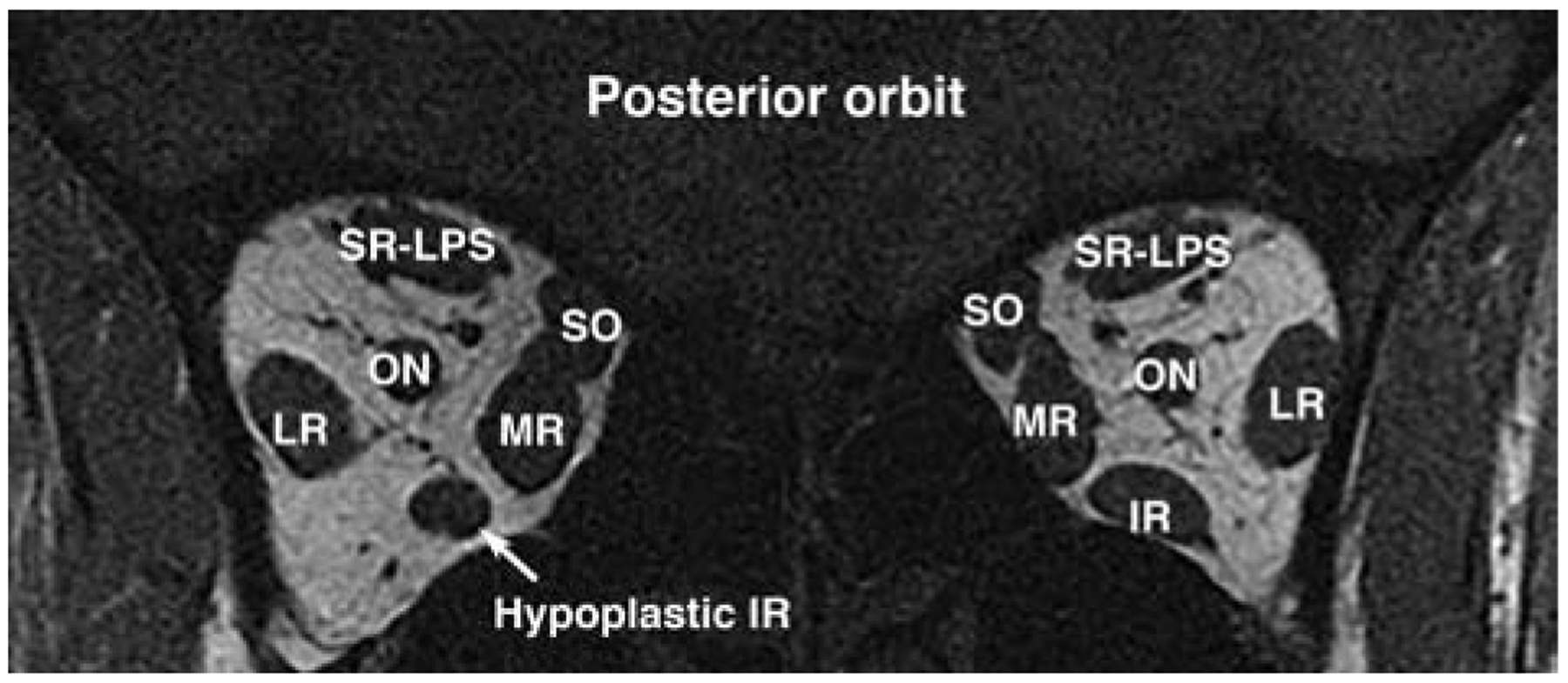
Background: Inferior rectus (IR) underaction may arise from various causes that are distinguishable through imaging. We investigated clinical and imaging characteristics of congenital and acquired causes of IR underaction.

Methods: Cases of IR underaction were selected from data prospectively collected in a study of orbital imaging in strabismic patients.

Results: Review identified 3 cases of congenital IR underaction (2 with bilateral IR aplasia and 1 with unilateral IR hypoplasia), 12 acquired cases, including 4 due to denervation (2 idiopathic, 1 after multiple strabismus surgeries, 1 after head trauma), and 8 cases of direct IR damage (5 with orbital trauma and 3 with previous surgery, including 2 sinus surgery and 1 laser blepharoplasty). Of the 23 cases, 11 adults had high-resolution magnetic resonance imaging, and 2 children had computed tomography. Imaging identified the anatomic diagnosis in congenital cases; in acquired cases, imaging helped to identify atrophy and exclude alternative orbital causes; and in direct mechanical damage, imaging clarified the mechanism of underaction, extent of IR damaged, and the degree of retained contractility. Patients with congenital IR absence or hypoplasia exhibited A pattern exotropia that was typically absent in isolated acquired denervation or direct IR damage.

Conclusions: Orbital imaging demonstrates a variety of abnormalities in patients with congenital or acquired IR hypofunction, helping to clarify the underlying mechanism and guide management.


Figures

FIG 1.
FIG 1.
Versions of case 1 with bilateral congenital absence of the inferior rectus muscle (IR), demonstrating limited infraduction bilaterally, especially in abduction, with A pattern exotropia reaching an estimated 100Δ with maximal infraducting effort not fully captured here.
FIG 2.
FIG 2.
Case 1. Quasi-coronal T2 MRI of the right (left column) and left (right column) orbits showing bilateral absence of the inferior rectus (IR), narrowing and hypoplasia of the superior rectus (SR), incyclorotation of rectus pulley array in the right more than in the left orbit, and an abnormal band from the SR to the inferior oblique (IO) in the right orbit. IR, inferior rectus; LPS, levator palpebrae superioris muscle; LR, lateral rectus; MR, medial rectus; N to IO, nerve to inferior oblique; ON, optic nerve; SO, superior oblique.
FIG 3.
FIG 3.
Surgical exposure of right orbit of case 1. A, Inferior sclera shows absence of IR muscle, tendon, and ciliary vessels. B, Abnormally narrowed SO tendon. C, A 6–0 polyglactin 910 suture was placed just posterior and lateral to the terminal portion of the SO insertion, and then passed through the anterior portion of the SO tendon at the insertion. The anterior three-quarters of the SO tendon were then divided posterior to the suture and detached. D, The suture was then tied to fold and rotate the anterior portion of the SO tendon posteriorly to the scleral suture site to enhance infraduction action. E, Diagram of step C, suturing the anterior tendon margin and posterior scleral pass before tenotomy. F, Diagram of step D: posterior folding of the anterior SO tendon. SR, superior rectus.
FIG 4.
FIG 4.
Case 2. Quasi-coronal T2 MRI of the orbits showing moderately hypoplastic right inferior rectus muscle. SR-LPS, superior rectus–levator palpebrae superioris complex.
FIG 5.
FIG 5.
Case 3. One mm thickness coronal CT scan demonstrate bilateral absence of the inferior rectus (IR).
FIG 6.
FIG 6.
Case 4. Sagittal (top row) and coronal (bottom row) MRI of right (left column) and left (right column) orbits in central gaze, demonstrating atrophic left IR with inferior bowing.
FIG 7.
FIG 7.
Case 5. Quasi-coronal T2 MRI of right (left column) and left (right column) orbits of case 1 in supraduction (top row), central gaze (middle row), and infraduction (bottom row) showing atrophy of the left IR that lacks contractile changes with attempted infraduction.
FIG 8.
FIG 8.
Case 8. Quasi-coronal of posterior, medium and anterior (left and middles columns) and sagittal (right column) T2 MRI of the left orbit in up (top row), primary (central row), and down gaze (bottom row) showing disorganization of the left intraocular content with total retinal detachment, general commotion to the extraocular muscles and an inferomedial fluid collection near the orbital implant. The anterior half of the left IR was not clearly visible. The posterior half of the IR has retained bulk with retained contractility.
FIG 9.
FIG 9.
Case 9. Sagittal T2 MRI showing disrupted connective tissue in the region of the IR pulleys. The anterior third of the IR exhibits a T2-weighted imaging signal (*) similar to that of the orbital fat, with normal signal more posteriorly and intact innervation.
FIG 10.
FIG 10.
Case 12. Coronal (left column) and sagittal (right column) T2 MRI in central (top and mid rows) and down gaze (bottom row) showing a functional IR in normal continuity with the scleral insertion surrounded by a large contrast enhanced mass (*). Right IR shows changes in attempted infraduction.

Similar articles

Cited by

References

    1. Chou TM, Demer JL. Isolated inferior rectus palsy caused by a metastasis to the oculomotor nucleus. Am J Ophthalmol 1998;126:737–40. - PubMed
    1. Akbari MR, Ameri A, Keshtkar Jaafari A, Fard MA, Eshraghi B, Mirmohammadsadeghi A. Clinical features and surgical outcomes of isolated inferior rectus muscle paralysis. Strabismus 2014;22:58–63. - PubMed
    1. Awadein A Clinical findings, orbital imaging, and intraoperative findings in patients with isolated inferior rectus muscle paresis or underaction. J AAPOS 2012;16:345–9. - PubMed
    1. Young SM, Koh YT, Chan EW, Amrith S. Incidence and risk factors of inferior rectus muscle palsy in pediatric orbital blowout fractures. Craniomaxillofac Trauma Reconstr 2018;11:28–34. - PMC - PubMed
    1. Khitri MR, Demer JL. Magnetic resonance imaging of tissues compatible with supernumerary extraocular muscles. Am J Ophthalmol 2010;150:925–31. - PMC - PubMed

Publication types