. 2021 Jun 2;29(6):2134-2150.
doi: 10.1016/j.ymthe.2021.02.017. Epub 2021 Feb 15.

WDR5 facilitates EMT and metastasis of CCA by increasing HIF-1α accumulation in Myc-dependent and independent pathways

Affiliations

WDR5 facilitates EMT and metastasis of CCA by increasing HIF-1α accumulation in Myc-dependent and independent pathways

Tianli Chen et al. Mol Ther. .

Abstract

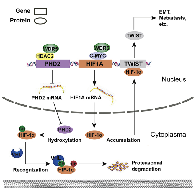
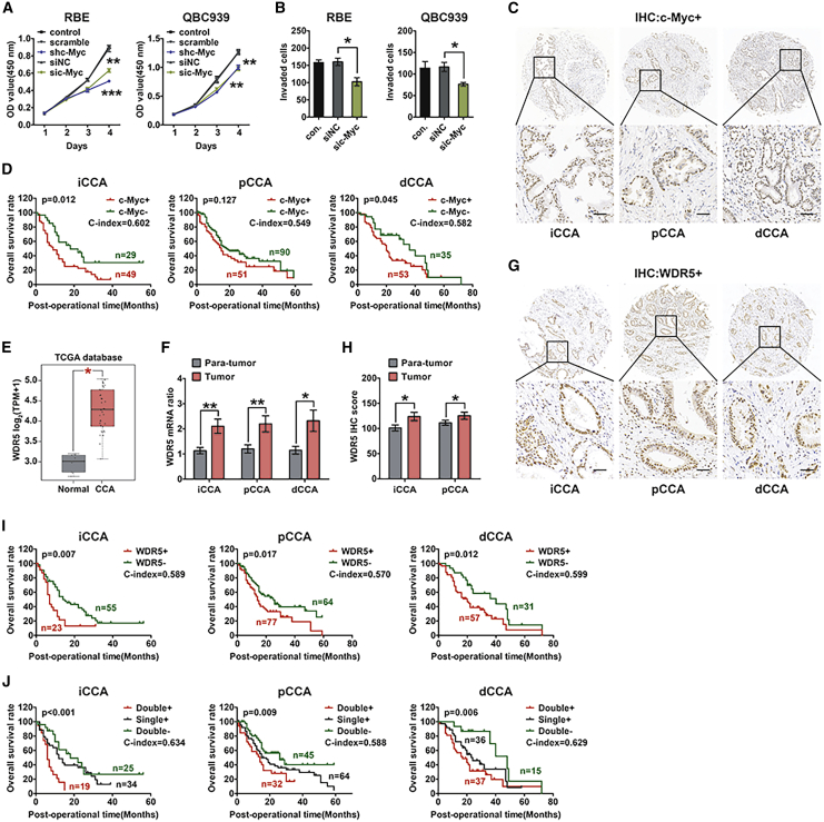
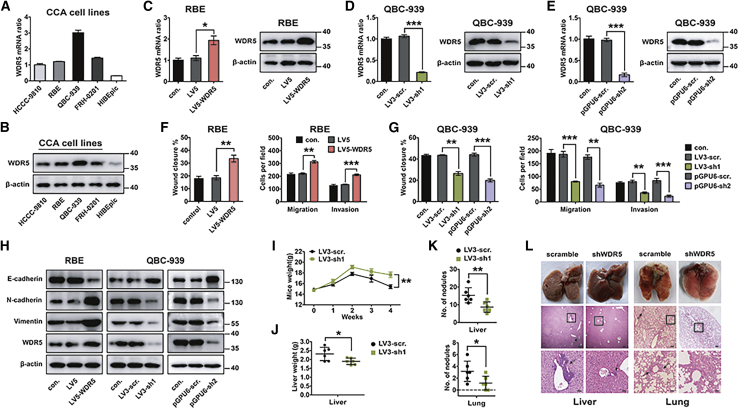
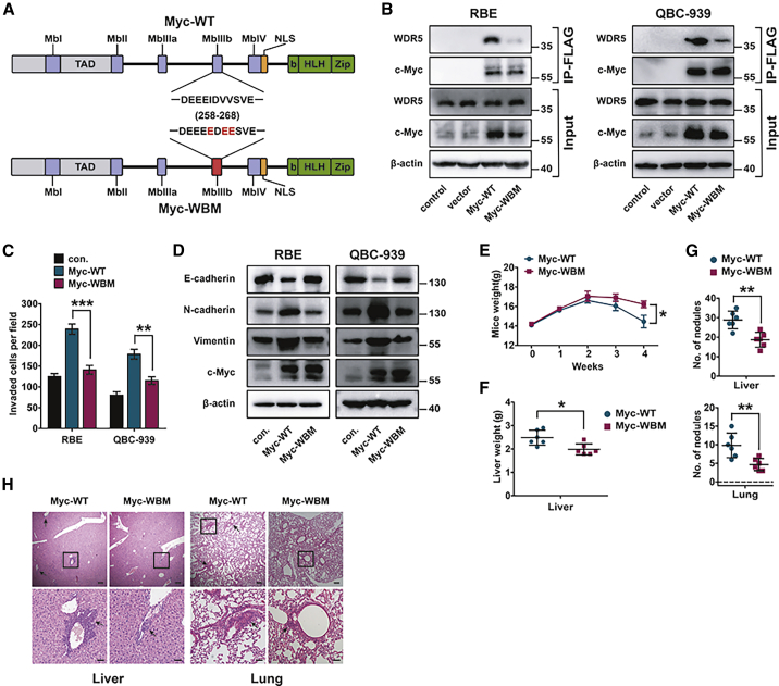
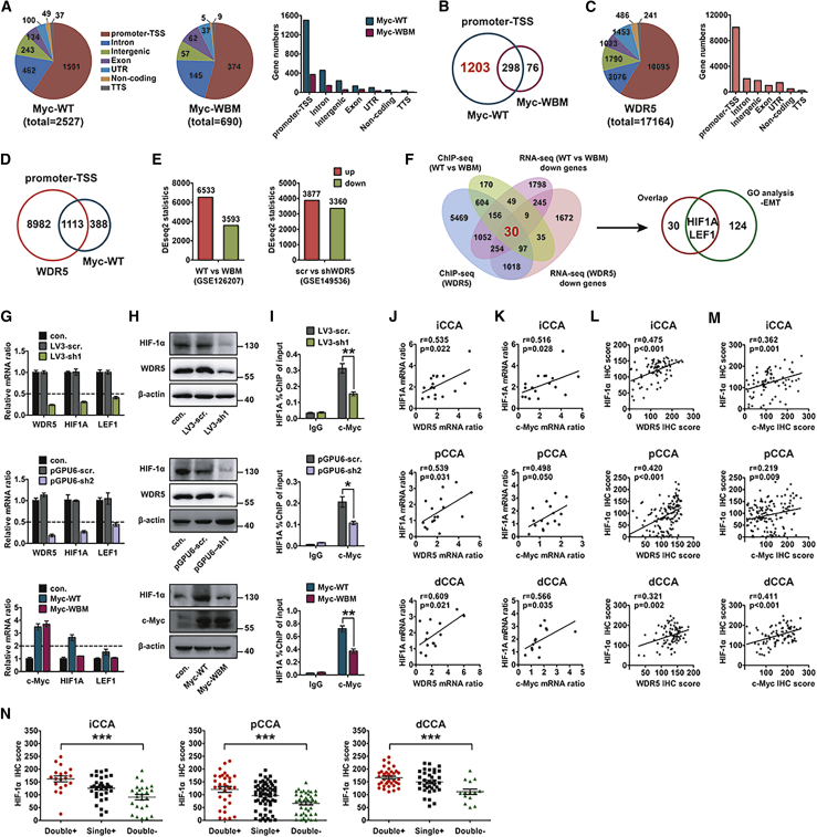
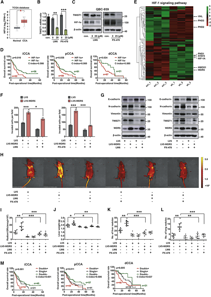
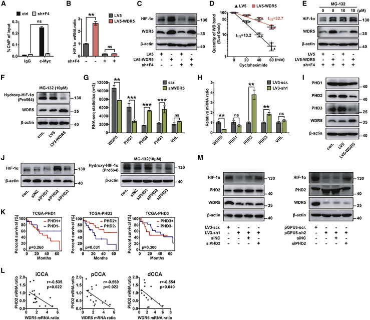
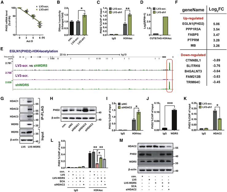
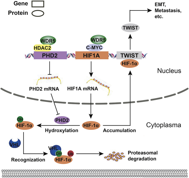
Cholangiocarcinoma (CCA) is a highly aggressive malignancy with extremely poor prognoses. The oncogenic role and prognostic value of c-Myc in CCA is not well elucidated. WD repeat domain 5 (WDR5) is a critical regulatory factor directly interacting with c-Myc to regulate c-Myc recruitment at chromosomal locations, but the interaction of WDR5 and c-Myc in CCA was uncovered. In our study, we detected WDR5 and c-Myc expression in all CCA types, including intrahepatic (iCCA), perihilar (pCCA), and distal (dCCA) CCA, and evaluated their prognostic significance. Consequently, we demonstrated that WDR5 was significantly correlated with poor prognosis of CCA and that WDR5 and c-Myc co-expression was a more sensitive prognostic factor. With in vitro and in vivo experiments and bioinformatics, we showed that WDR5 interacted with the Myc box IIIb (MBIIIb) motif of c-Myc and facilitated Myc-induced HIF1A transcription, thereby promoting the epithelial-mesenchymal transition (EMT), invasion, and metastasis of CCA. Moreover, WDR5 enhanced hypoxia-inducible factor 1 subunit α (HIF-1α) accumulation by binding with histone deacetylase 2 (HDAC2) and increasing histone 3 lysine 4 acetylation (H3K4ac) deacetylation of the prolyl hydroxylase domain protein 2 (PHD2) promoter, resulting in the attenuation of chromatin opening and PHD2 expression, and eventually leading to HIF-1α stabilization and accumulation. In conclusion, WDR5 facilitated EMT and metastasis of CCA by increasing HIF-1α accumulation in a Myc-dependent pathway to promote HIF-1α transcription and a Myc-independent pathway to stabilize HIF-1α.

Keywords: CCA; EMT; HIF-1α; PHD2; WDR5; c-Myc.


Conflict of interest statement

Declaration of interests The authors declare no competing interests.

Figures

None
Graphical abstract
Figure 1
Figure 1
Co-expression of high c-Myc and WDR5 was correlated with the poorest prognosis of CCA (A and B) Knockdown of c-Myc inhibited cell proliferation (A) and invasion (B) of the iCCA cell line RBE and the pCCA cell line QBC939. (C) Representative IHC staining of c-Myc in iCCA, pCCA, and dCCA. Scale bars, 50 μm. (D) Overall survival curves of iCCA, pCCA, and dCCA patients were stratified by c-Myc expression. (E) TCGA data of WDR5 mRNA level in 9 normal tissues and 36 CCA tissues. The expression of WDR5 mRNA was quantified using log2(TPM + 1). (F) mRNA levels of WDR5 in 18 iCCA, 16 pCCA, and 14 dCCA tissues as well as their corresponding normal tissues. (G) Representative IHC staining of WDR5 expression in iCCA, pCCA, and dCCA tissues. (H) IHC scores of WDR5 in iCCA, pCCA, and corresponding normal tissues. (I) Overall survival curves of iCCA, pCCA, and dCCA patients were stratified by WDR5 expression. (J) Survival curves were further stratified into subgroups with the co-expression, single expression, and low expression of c-Myc and WDR5. ∗p < 0.05, ∗∗p < 0.01. In (A), data were calculated by two-way ANOVA. In (B) and (E), data were calculated by one-way ANOVA. In (F) and (H), data were calculated by a paired t test. In (D), (I), and (J), data were calculated by a log-rank test, and the C-index was calculated to evaluate the accuracy of the prognostic model.
Figure 2
Figure 2
WDR5 facilitated the migration, invasion, and metastasis of CCA (A and B) The expression of WDR5 in different CCA cell lines and in a normal biliary epithelium cell line (HIBEpiCs) was detected with qPCR (A) and western blot (B). (C) WDR5 was overexpressed in RBE cells with lentivirus encoding LV5-WDR5. (D and E) WDR5 was knocked down in QBC939 cells with two different shRNAs in different vectors, namely LV3-shWDR5-1 (D) and pGPU6-shWDR5-2 (E). (F and G) Invasion and migration of RBE (F) and QBC939 (G) cells were detected with wound-healing (left panel) and transwell assays (right panel) after overexpressing or silencing WDR5. (H) EMT biomarkers were detected after WDR5 overexpression in RBE cells and knockdown in QBC939 cells. (I) Stable WDR5-silenced cells and control cells were injected via the tail vein of BALB/c nude mice. The body weights of mice injected with scramble (LV3-scr.) and shWDR5 (LV3-sh1) cells were measured weekly. (J) Livers were weighed to assess the tumor burden of liver metastatic foci. (K) Number of metastasis nodules in livers and lungs of mice. (L) Representative gross specimens and H&E staining of metastasis lesions in liver and lung. Arrows indicate the metastasis lesions. Scale bars, 50 μm. ∗p < 0.05, ∗∗p < 0.01, ∗∗∗ p < 0.001. In (C)–(K), data were analyzed with a one-way ANOVA. In (I), data were analyzed with a two-way ANOVA.
Figure 3
Figure 3
WDR5 promoted CCA metastasis via the interaction with c-Myc (A) Schematic diagram of human wild-type (WT) and WBM mutant c-Myc protein. The MBIIIb motif refers to amino acids from 258 to 268. WBM represents the I262E/V264E/V265E mutation of the MBIIIb motif. (B) Co-immunoprecipitation (coIP) of WT and WBM mutant c-Myc with endogenous WDR5 in RBE (left) and QBC939 (right) cells. (C) The invasion of RBE and QBC939 cells transfected with Myc-WT or Myc-WBM was detected with a transwell assay. (D) In RBE and QBC939 cells stably expressing Myc-WT or Myc-WBM, the expression levels of EMT-specific proteins were detected with western blot. (E) Weekly body weight of BALB/c mice injected with QBC939 cells overexpressing Myc-WT or Myc-WBM. (F) Livers were weighed to assess the tumor burden of liver metastatic foci. (G) Number of metastasis nodules in livers and lungs of mice. (H) Representative H&E staining of metastasis lesions in liver and lung. ∗p < 0.05, ∗∗p < 0.01, ∗∗∗p < 0.001, calculated with a two-way ANOVA (E) or with a one-way ANOVA (C, F, and G).
Figure 4
Figure 4
HIF-1α was the primary direct target of c-Myc and WDR5 (A) Re-analysis of the ChIP-seq datasets regarding Myc-WT and Myc-WBM in HEK293 cells (GEO: GSE60897). Pie chart and histogram show the percentage and number of different binding regions mapped for WT and WBM of c-Myc. (B) Venn diagram shows the overlap of genes binding with Myc-WT and Myc-WBM. (C) ChIP-seq datasets show the number and binding sites of genes interacting with WDR5 in HEK293 cells (GEO: GSE60897). (D) Overlapped genes interacting with the promoter-TSS region of both WDR5 and Myc-WT are shown in the Venn diagram. (E) Left panel: mutual regulated mRNAs in Ramos cells, a lymphoma cell line, with overexpression of Myc-WT and Myc-WBM (GEO: GSE126207). Right panel: WDR5 in QBC939 cells was silenced by shRNA, and mRNA sequencing was performed to screen the upregulated or downregulated genes (GEO: GSE149536). (F) Flowchart of screening downstream genes of c-Myc and WDR5 by the bioinformatics method. Upper panel: a total of 30 overlapped genes were screened out by the results in the ChIP-seq datasets and RNA-seq, which are shown in (A)–(E). Bottom panel: the overlapped 30 genes had only 2 common genes with EMT-involved genes in GO analysis, which were HIF1A and LEF1. (G) In QBC939 cells, mRNAs of HIF1A and LEF1 were detected with qPCR after silencing WDR5 (upper two panels) or overexpressing Myc-WT/WBM (bottom panel). (H) HIF-1α expression was detected with western blot after silencing WDR5 (upper two panels) or overexpressing Myc-WT/WBM (bottom panel) in QBC939 cells. (I) ChIP assay revealed that WDR5 knockdown (upper two panels) or Myc-WBM mutation (bottom panel) inhibited the recruitment of c-Myc on the HIF1A promoter. ∗p < 0.05, ∗∗p < 0.01, by one-way ANOVA. (J and K) mRNA correlations between HIF-1α and WDR5 (J) or c-Myc (K) were analyzed with a Pearson correlation test. (L and M) Protein correlations between HIF-1α and WDR5 (L) or c-Myc (M) were analyzed with a Pearson correlation test. (N) Protein levels of HIF-1α in CCA patients with double-positive, single-positive, and double-negative expression of c-Myc and WDR5. ∗∗∗p < 0.001, by one-way ANOVA.
Figure 5
Figure 5
HIF-1α was an important effector in WDR5-induced CCA metastasis (A) HIF1A mRNA in CCAs was significantly higher than that in normal tissues. (B and C) Quantitative real-time PCR (B) and western blot (C) show that the HIF-1α inhibitor LW6 (20 μM) or PX-478 (25 μM) decreased TWIST1 expression. (D) Survival curves of iCCA, pCCA, and dCCA patients were stratified by HIF-1α expression. (E) Genes in the HIF1A signaling pathway were changed along with WDR5 knockdown. (F and G) Invasive ability (F) and expression of EMT-specific proteins (G) in WDR5-overexpressed QBC939 cells in the presence or absence of LW6 (20 μM) or PX-478 (25 μM). (H) Metastatic models were established by tail vein injection of WDR5-overexpressed QBC939 cells in the treatment of HIF-1α inhibitor. Two HIF-1α inhibitors, LW6 (10 mg/kg p.o.) and PX-478 (100 mg/kg i.p.), were used to inhibit HIF-1α expression in vivo. The tumor metastases were monitored by a live imaging system (n = 6). (I) Radiant efficiency of in vivo fluorescence in (H) was measured to quantify the tumor burden of mice. (J) Livers of mice in (H) were weighed to assess the tumor burden of liver metastatic foci. (K and L) The numbers of metastasis nodules in livers (J) and lungs (K) of mice in (H) were counted. (M) The survival curves were further stratified into subgroups with the co-expression, single expression, and double low expression of WDR5 and HIF-1α in iCCA, pCCA and dCCA. In (B), (F), and (I)–(L), ∗p < 0.05, ∗∗p < 0.01, ∗∗∗p < 0.001, calculated by one-way ANOVA. In (D) and (M), data were calculated by a log-rank test, and the C-index was calculated to evaluate the accuracy of the prognostic model.
Figure 6
Figure 6
WDR5 promoted HIF-1α accumulation via suppressing the PHD2-mediated degradation pathway (A) ChIP assay proved that the combination of shMyc (sh) and c-Myc inhibitor 10058-F4 (40 μM) almost completely suppressed c-Myc recruitment on the HIF1A promoter. (B and C) In QBC939 cells treated with shMyc and inhibitor 10058-F4, quantitative real-time PCR (B) and western blot (C) showed that WDR5 overexpression had little effect on HIF1A mRNA, but it increased HIF-1α accumulation. (D) Half-life analysis of HIF-1α protein. Cells were treated with 10 μg/mL cycloheximide (CHX) for the indicated times. The gray scale values of western blot bands were quantified with ImageJ software. (E) Effect of ubiquitination inhibitor MG-132 (10 μM) on control and WDR5-overexpressed QBC939 cells. MG-132 attenuated the HIF-1α elevation induced by WDR5 overexpression in QBC939 cells treated with shMyc and inhibitor 10058-F4. (F) In QBC939 cells treated with shMyc and inhibitor 10058-F4, WDR5 overexpression decreased hydroxylation of HIF-1α. (G and H) The mRNA levels of the PHD family and VHL in QBC939 cells transfected with scramble and WDR5 knockdown were detected by RNA-seq (G) and verified by quantitative real-time PCR (H). (I) The expression levels of the PHD family in QBC939 cells transfected with control, vector, and LV5-WDR5 were detected by western blot in the treatment with shMyc and inhibitor 10058-F4. (J) Effect of PHD1–PHD3 knockdown on the expression of total HIF-1α and hydroxy-HIF-1α in QBC939 cells treated with shMyc and inhibitor 10058-F4. (K) Survival curves of CCA were stratified with PHD1–PHD3 in TGCA database. (L) The correlations between WDR5 and PHD2 mRNA in iCCA, pCCA, and dCCA were analyzed with a Pearson correlation test. (M) Expression levels of HIF-1α in WDR5-silenced and/or PHD2-silenced QBC939 cells after treatment with shMyc and inhibitor 10058-F4. PHD2 knockdown attenuated the influence of WDR5 knockdown on HIF-1α reduction. Data were analyzed with one-way ANOVA in (A), (B), and (H). ∗p < 0.05, ∗∗p < 0.01, ∗∗∗p < 0.001. n.s., not significant.
Figure 7
Figure 7
WDR5 suppressed PHD2 expression through HDAC2-induced H3K4 deacetylation (A) Half-life detection of PHD2 mRNA was performed using 10 μM actinomycin D (Act D) for the indicated times. (B) Chromatin accessibility of PHD2 promoters was evaluated by quantitative real-time PCR with DNase I-pretreated nucleus of control, scramble, and WDR5-silenced QBC939 cells. Fold change was analyzed using 2−ΔΔCt. (C) ChIP assay showed that WDR5-silenced QBC939 cells had elevated H3K4ac modification at the PHD2 promoter regions. H3K4ac antibody was used for immunoprecipitation (IP) after WDR5 knockdown, and quantitative real-time PCR of the PHD2 promoter in output was performed. (D) A CUT&Tag assay was performed to confirm that WDR5 knockdown promoted H3K4ac modification at PHD2 promoter regions. WDR5 was knocked down by shWDR5 in QBC939 cells, and H3K4Ac antibody was used to interact with the H3K4-acetylized protein. Log2(RPM + 1) was used for quantification. (E) Overlay of H3K4ac at EGLN1 (PHD2) loci in scramble (purple) and WDR5 knockdown (green) QBC939 cells. Significantly increased regions are underscored (red box). (F) List of 10 DEGs with most significant change in CUT&Tag data. EGLN1 (PHD2) had the highest fold change after WDR5 knockdown. (G) CoIP of WDR5 with HDAC1–HDAC3 in QBC939 cells. (H) Effect of HDAC1–HDAC3 knockdown on the expression of PHD2 protein in QBC939 cells. (I) ChIP assays show that H3K4ac on PHD2 promoters in HDAC2 silenced QBC939 cells. H3K4ac antibody was used for IP after HDAC2 knockdown, and qPCR of the PHD2 promoter in output was performed. (J) ChIP assay revealed that WDR5 directly interacts with the promoter region of PHD2 gene. WDR5 antibody was used for IP, and qPCR of the PHD2 promoter in output was performed. (K) ChIP assay revealed that WDR5 knockdown attenuated HDAC2 binding to the PHD2 promoter. HDAC2 antibody was used for IP after WDR5 knockdown. (L) ChIP assay revealed that WDR5 overexpression disrupted H3K4ac at PHD2 promoter regions and knockdown or inhibition of HDAC2 reversed it. H3K4ac antibody was used for IP, and quantitative real-time PCR of PHD2 promoter in output was performed. (M) Expression of PHD2 in WDR5-overexpressed QBC939 cells that were treated with 50 μM SCA for 48 h or transfected with siHDAC2. Data were analyzed with two-way ANOVA in (A), or with one-way ANOVA in (B), (C), and (I)–(L). ∗p < 0.05, ∗∗p < 0.01, ∗∗∗p < 0.001. n.s., not significant.
Figure 8
Figure 8
Schematic depiction of the mechanisms underlying WDR5 facilitating CCA metastasis and EMT by increasing HIF-1α accumulation Two distinct mechanisms of how WDR5 enhanced HIF-1α accumulation: (1) WDR5 promoted HIF-1α mRNA synthesis via directly interacting with c-Myc and (2) WDR5 inhibited HIF-1α protein hydroxylation, ubiquitination, and degradation through suppressing PHD2 expression, which relied on interacting with HDAC2 and deacetylizing H3K4ac.

Similar articles

Cited by

References

    1. Rizvi S., Khan S.A., Hallemeier C.L., Kelley R.K., Gores G.J. Cholangiocarcinoma—evolving concepts and therapeutic strategies. Nat. Rev. Clin. Oncol. 2018;15:95–111. - PMC - PubMed
    1. Pitt H.A., Nakeeb A., Abrams R.A., Coleman J., Piantadosi S., Yeo C.J., Lillemore K.D., Cameron J.L. Perihilar cholangiocarcinoma. Postoperative radiotherapy does not improve survival. Ann. Surg. 1995;221:788–797. discussion 797–798. - PMC - PubMed
    1. Xu Y.F., Yang X.Q., Lu X.F., Guo S., Liu Y., Iqbal M., Ning S.L., Yang H., Suo N., Chen Y.X. Fibroblast growth factor receptor 4 promotes progression and correlates to poor prognosis in cholangiocarcinoma. Biochem. Biophys. Res. Commun. 2014;446:54–60. - PubMed
    1. Blechacz B. Cholangiocarcinoma: current knowledge and new developments. Gut Liver. 2017;11:13–26. - PMC - PubMed
    1. DeOliveira M.L., Cunningham S.C., Cameron J.L., Kamangar F., Winter J.M., Lillemoe K.D., Choti M.A., Yeo C.J., Schulick R.D. Cholangiocarcinoma: thirty-one-year experience with 564 patients at a single institution. Ann. Surg. 2007;245:755–762. - PMC - PubMed

Publication types

MeSH terms

Substances