

Clinical and Radiological Follow-Up Results of Patients with Sequestered Lumbar Disc Herniation: A Prospective Cohort Study
- PMID: 33601393
- PMCID: PMC8280406
- DOI: 10.1159/000515308
Clinical and Radiological Follow-Up Results of Patients with Sequestered Lumbar Disc Herniation: A Prospective Cohort Study
Abstract
Purpose: The aim of the study was to assess radiological changes and clinical outcomes of patients with sequestered lumbar disc herniation (LDH) and evaluate the relationship between them.
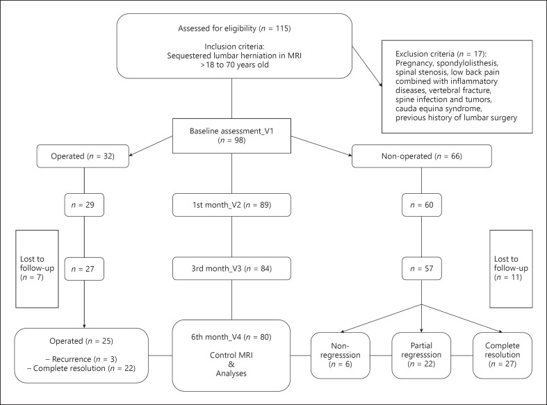
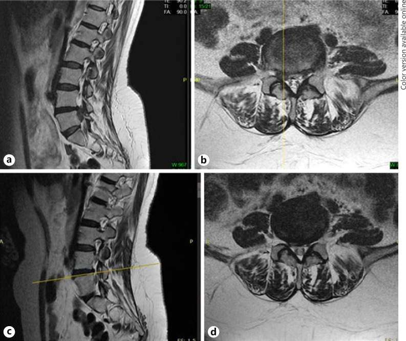
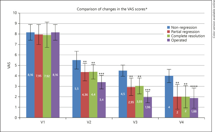
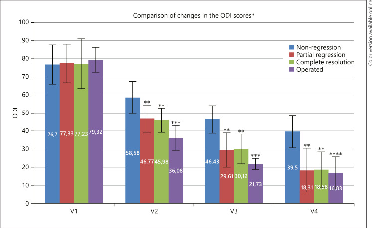
Methods: Patients diagnosed with sequestered LDH were followed up in 2 groups: operated (within the 1st month after diagnosis) and nonoperated. Visual analog scale (VAS) and Oswestry Disability Index (ODI) scores at baseline (V1) and 1st (V2), 3rd (V3), and 6th (V4) month visits were used for clinical evaluation. Radiological evaluation was performed by measuring the sequestered LDH level and herniation volume using magnetic resonance imaging (MRI) at V1 and V4. After the second MRI, patients in the nonoperated group were divided into 3 groups: nonregression (n = 6), partial regression (n = 22), and complete resolution (n = 27); patients were analyzed in 4 groups including the ones in the operated (n = 25) group.
Results: Significant improvements were observed in VAS and ODI scores at V2 and V3 in all groups (p = 0.000) and at V4 in partial regression and complete resolution groups (p = 0.000). VAS and ODI score improvements at V2 and V3 were significantly higher in the operated group than in other groups (p = 0.000). At V4, there were no significant differences in VAS and ODI scores (p > 0.05) between the operated group and partial regression and complete resolution groups.
Conclusion: Spontaneous regression was observed in the 6th month post-MRI in most of the nonoperated sequestered LDH patients with conservative treatment. Improvements in pain and disability scores were higher among the operated patients at the early stage, whereas they were not significantly different compared to patients with spontaneous regression at the 6th month.
Keywords: Lumbar disc herniation; Magnetic resonance imaging; Neurology; Pain; Radiology; Rehabilitation; Sequestered lumbar disc herniation; Spontaneous regression.
© 2021 The Author(s) Published by S. Karger AG, Basel.
Conflict of interest statement
The authors declare that they have no conflict of interest.
Figures
References
-
- Fardon DF, Milette PC. Nomenclature and classification of lumbar disc pathology. Recommendations of the Combined Task Forces of the North American Spine Society, American Society of Spine Radiology, and American Society of Neuroradiology. Spine. 2001;26((5)):E93–113. - PubMed
-
- Albert HB, Manniche C. The efficacy of systematic active conservative treatment for patients with severe sciatica: a single-blind, randomized, clinical, controlled trial. Spine. 2012;37((7)):531–42. - PubMed
-
- Ahn SH, Park HW, Byun WM, Ahn MW, Bae JH, Jang SH, et al. Comparison of clinical outcomes and natural morphologic changes between sequestered and large central extruded disc herniations. Yonsei Med J. 2002;43((3)):283–90. - PubMed
MeSH terms
LinkOut - more resources
Full Text Sources
Other Literature Sources
Medical
Research Materials
