

Comment
doi: 10.1084/jem.20202071.
Gatekeepers of the fetus: Characterization of placental macrophages
Affiliations
- PMID: 33601417
- PMCID: PMC7754679
- DOI: 10.1084/jem.20202071
Item in Clipboard
Comment
Gatekeepers of the fetus: Characterization of placental macrophages
J Exp Med.
.
Abstract
In this issue of JEM, Thomas et al. (https://doi.org/10.1084/jem.20200891) provide elegant technological and conceptual advances that further our understanding of the immune cells enriched at the maternal-fetal interface. Using new isolation strategies to better separate maternal- and fetal-derived cells, the authors identify previously undefined maternal-derived immune cells associated with the fetal-derived placenta and provide an in-depth analysis of the markers and characteristics of placental Hofbauer cells.
© 2020 Megli and Coyne.
Figures

Insights from Christina Megli and Carolyn B. Coyne
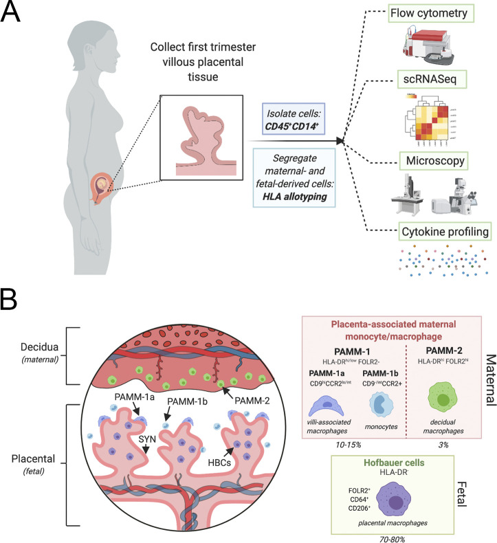
Isolation and characterization of placental associated macrophages and monocytes. (A) Cells were isolated from first trimester placental explants. CD14/CD45+ cells stained for HLA allotype to distinguish between maternal- and fetal-derived cells were sorted through a flow cytometer and characterized by transcriptional profile, microscopic evaluation, and staining as well as cytokine profiling to identify and characterize the distinct maternal and fetal macrophages in the placenta. (B) Left: The maternal–fetal interface is composed of the maternal-derived decidua and fetal-derived placenta. In the first trimester, chorionic villi are immature and are covered by a contiguous layer called the syncytiotrophoblast (dark pink, SYN). Thomas et al. (2020) define the characteristics of both maternal- and fetal-derived macrophages, termed PAMMs and HBCs. PAMM-1 (dark blue and light blue) and PAMM-2 (green) cells are identified by their HLA-DR, FOLR2 surface expression. PAMM-1 subtypes are distinct monocytes (PAMM-1b, light blue) and macrophages (PAMM-1a, dark blue). PAMM-1b macrophages are in direct contact with the SYN layer. The fetal-derived HBCs are in direct contact with the stroma of the chorionic villi and make up the majority of CD14+ cells in the placenta (70–80%), whereas PAMM-1 and PAMM-2 cells make up ∼10% and 3%, respectively. These cells are able to be identified by HLA-DR, FOLR2, CD64, and CD206 and are functionally and transcriptionally distinct from the maternal macrophage subsets. This figure was constructed using BioRender.
Comment on
-
Phenotypic and functional characterization of first-trimester human placental macrophages, Hofbauer cells.J Exp Med. 2021 Jan 4;218(1):e20200891. doi: 10.1084/jem.20200891. J Exp Med. 2021. PMID: 33075123 Free PMC article. Clinical Trial.
References
Publication types
MeSH terms
Grants and funding
LinkOut - more resources
Full Text Sources
Other Literature Sources
