. 2021 May 4;143(18):1797-1808.
doi: 10.1161/CIRCULATIONAHA.120.052671. Epub 2021 Feb 19.

Short-Chain Fatty Acids Outpace Ketone Oxidation in the Failing Heart

Affiliations

Short-Chain Fatty Acids Outpace Ketone Oxidation in the Failing Heart

Andrew N Carley et al. Circulation. .

Abstract

Background: The failing heart is energy starved with impaired oxidation of long-chain fatty acids (LCFAs) at the level of reduced CPT1 (carnitine palmitoyltransferase 1) activity at the outer mitochondrial membrane. Recent work shows elevated ketone oxidation in failing hearts as an alternate carbon source for oxidative ATP generation. We hypothesized that another short-chain carbon source, short-chain fatty acids (SCFAs) that bypass carnitine palmitoyltransferase 1, could similarly support energy production in failing hearts.

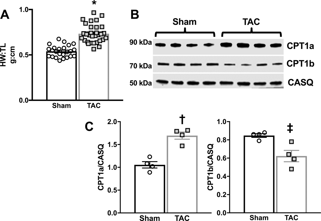
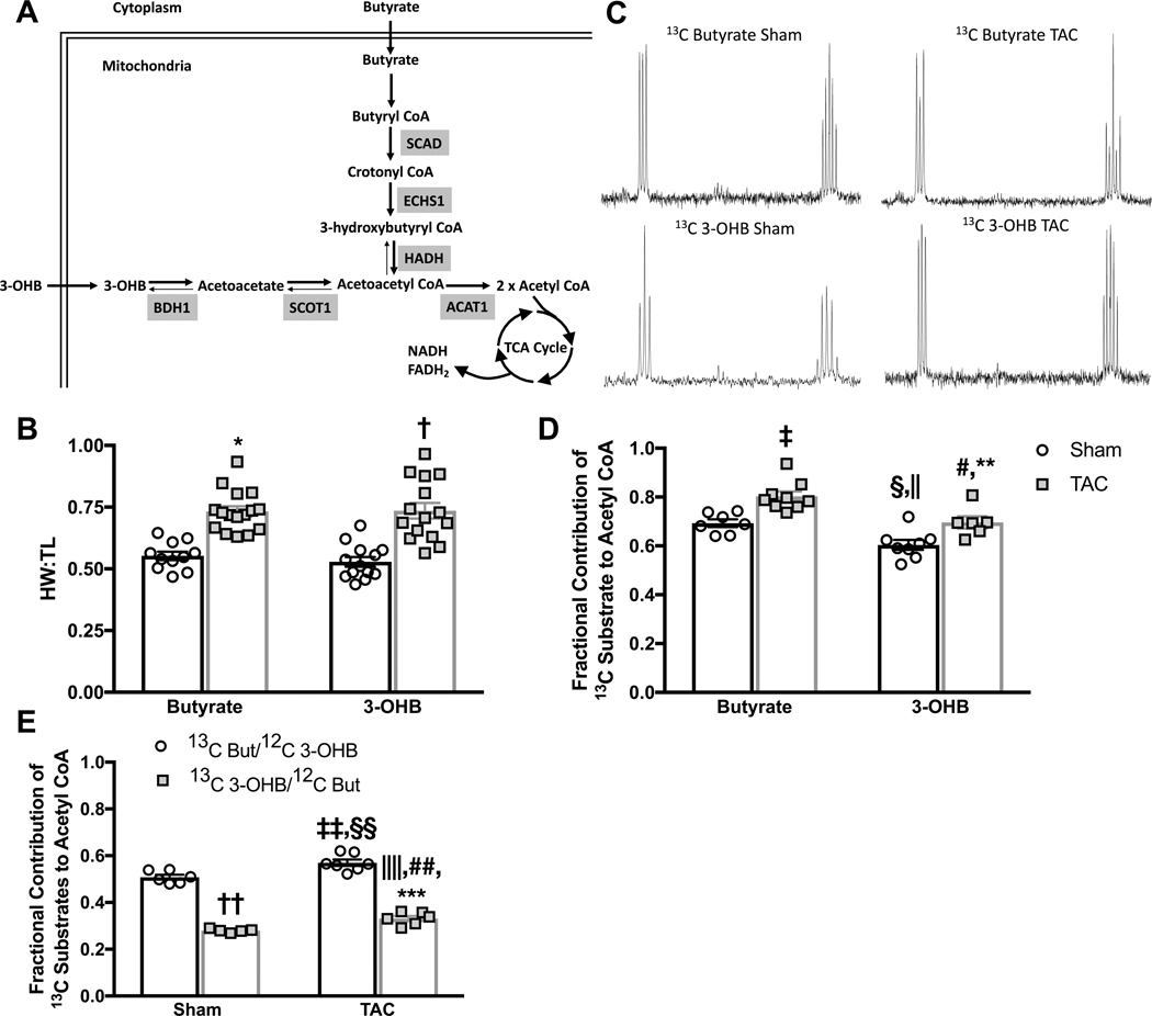
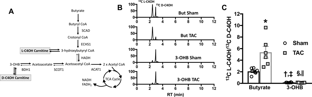
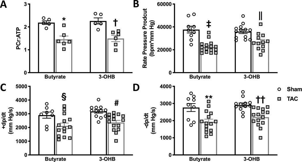
Methods: Cardiac hypertrophy and dysfunction were induced in rats by transverse-aortic constriction (TAC). Fourteen weeks after TAC or sham operation, isolated hearts were perfused with either the 4 carbon, 13C-labeled ketone (D3-hydroxybutyrate) or the 4 carbon, 13C-labeled SCFA butyrate in the presence of glucose and the LCFA palmitate. Oxidation of ketone and SCFA was compared by in vitro 13C nuclear magnetic resonance spectroscopy, as was the capacity for short-chain carbon sources to compensate for impaired LCFA oxidation in the hypertrophic heart. Adaptive changes in enzyme expression and content for the distinct pathways of ketone and SCFA oxidation were examined in both failing rat and human hearts.

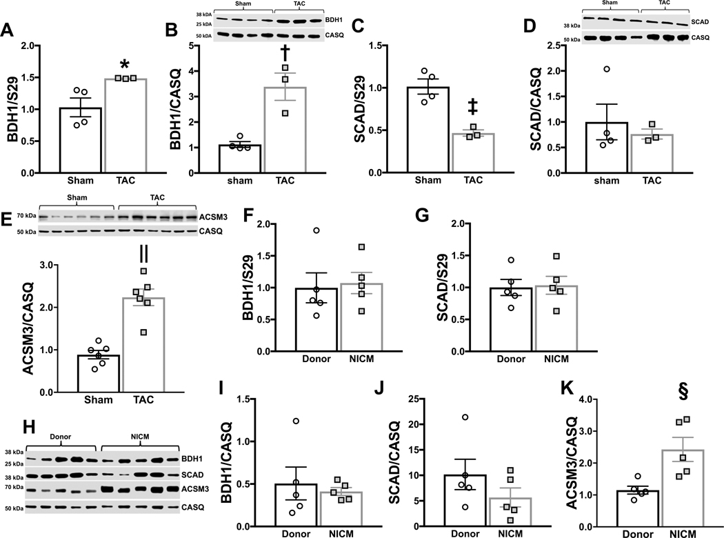
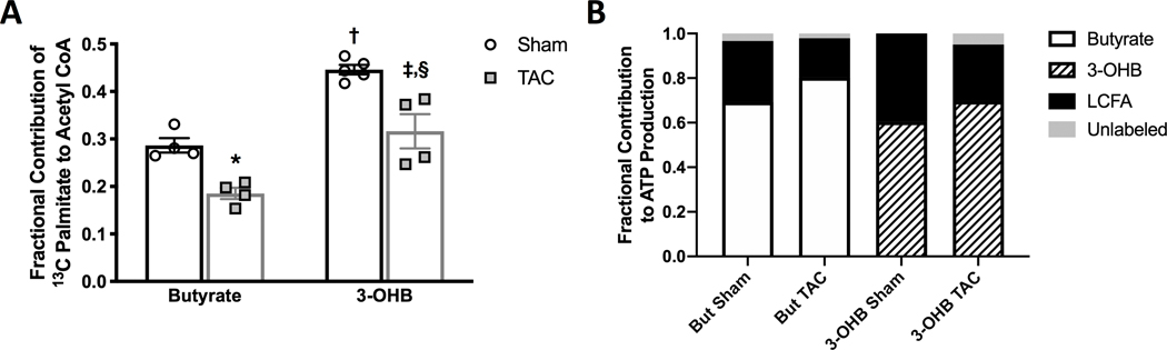
Results: TAC produced pathological hypertrophy and increased the fractional contributions of ketone to acetyl coenzyme-A production in the tricarboxylic acid cycle (0.60±0.02 sham ketone versus 0.70±0.02 TAC ketone; P<0.05). However, butyrate oxidation in failing hearts was 15% greater (0.803±0.020 TAC SCFA) than ketone oxidation. SCFA was also more readily oxidized than ketone in sham hearts by 15% (0.693±0.020 sham SCFA). Despite greater SFCA oxidation, TAC did not change short-chain acyl coenzyme-A dehydrogenase content. However, failing hearts of humans and the rat model both contain significant increases in acyl coenzyme-A synthetase medium-chain 3 enzyme gene expression and protein content. The increased oxidation of SCFA and ketones occurred at the expense of LCFA oxidation, with LCFA contributing less to acetyl coenzyme-A production in failing hearts perfused with SCFA (0.190±0.012 TAC SCFA versus 0.3163±0.0360 TAC ketone).

Conclusions: SCFAs are more readily oxidized than ketones in failing hearts, despite both bypassing reduced CPT1 activity and represent an unexplored carbon source for energy production in failing hearts.

Keywords: cardiac; heart failure; ketones; metabolism; short-chain fatty acid.


Figures

Figure 1 –
Figure 1 –. Hypertrophic response and carnitine palmitoyl transferase 1 expression in response to transverse aortic constriction (TAC).
A, Heart weight to tibia length (HW:TL) was measured in sham (n=24) and TAC (n=31) hearts 14 weeks after sham or TAC surgery; *p<0.0001 as determined by unpaired 2-tailed t-test. B, Carnitine palmitoyl transferase (CPT)1a and CPT1b expression in unperfused sham or TAC hearts isolated 14 weeks after TAC or sham surgery. C, CPT1a and CPT1b protein expression normalized to calsequestrin (CASQ); †p=0.0009, ‡p<0.0137 vs. sham as determined by unpaired 2-tailed t-test.
Figure 2 –
Figure 2 –. Fractional contribution of butyrate and 3-hydroxybutyrate (3-OHB) to mitochondrial oxidation.
A, model depicting the pathways for butyrate and 3-OHB oxidation in the heart. The role of enzymes acetyl CoA acetyltransferase (ACAT) 1, β-hydroxybutyrate dehydrogenase (BDH) 1, enoyl CoA hydratase short chain (ECHS) 1, 3-hydroxyacyl CoA dehydrogenase (HADH), short chain acyl CoA dehydrogenase (SCAD), and succinyl CoA:3-ketoacid CoA transferase (SCOT) 1 in short chain carbon metabolism is depicted. B, heart weight to tibia length (HW:TL) as measured in isolated hearts at the end of perfusion with either butyrate (n=11 sham, n=16 TAC) or 3-OHB (n=13 sham, n=15 TAC) perfused hearts; *p<0.0001 vs. butyrate sham, †p=0.0001 vs. 3-OHB sham group via 2-way ANOVA and Tukey’s post hoc test. C, representative spectra from end-point enrichment analysis of glutamate 13C enrichment in perfused hearts. D, fractional contribution of either 13C butyrate (n=7 sham, n=9 TAC) or 13C 3-OHB (n=8 sham, n=6 TAC) to acetyl CoA; ‡p=0.0032 vs. butyrate sham, §p=0.024 vs. butyrate sham, ||p<0.0001 vs. butyrate TAC, #p=0.0065 vs. butyrate TAC, **p=0.0252 vs. 3-OHB sham via 2-way ANOVA and Tukey’s post hoc test. E, fractional contribution of either 13C butyrate (n=6 sham, n=5 TAC) or 13C 3-OHB (n=7 sham, n=6 TAC) to acetyl CoA when provided as a 50:50 mix, ††p<0.0001 vs. 13C But/12C 3-OHB sham, ‡‡p=0.0034 vs. 13C But/12C 3-OHB sham, §§p<0.0001 vs. 13C 3-OHB/12C But sham, ||||p<0.0001 vs 13C But/12C 3-OHB sham, ##p=0.0273 vs. 13C 3-OHB/12C But sham, ***p<0.0001 vs. 13C But/12C 3-OHB TAC, via 2-way ANOVA and Tukey’s post hoc test
Figure 3 –
Figure 3 –. Expression of key enzymes regulating short chain fatty acid (SCFA) and ketone oxidation in the heart.
A, mRNA expression normalized to S29 of β-hydroxybutyrate dehydrogenase (BDH) 1, *p=0.0475 via two-tailed t-test. B, BDH1 protein expression normalized to calsequestrin (CASQ), †p=0.0047 via two-tailed t-test. C, mRNA expression normalized to S29 of short chain acyl CoA dehydrogenase (SCAD); ‡p=0.0041 via two-tailed t-test. D, SCAD protein expression normalized to CASQ. E, ACSM3 protein expression normalized to CASQ, ||p=0.0001 via two-tailed t-test. mRNA expression normalized to S29 for BDH 1 (F) and SCAD (G) from human donor hearts or hearts from patients with non-ischemic cardiomyopathy (NICM). H, protein expression for BDH1, SCAD and ACSM3 measured in donor hearts or patients with NICM. Protein expression for BDH1 (I), SCAD (J), and ACSM3 (K) normalized to CASQ, §p=0.0125 via two-tailed t-test.
Figure 4 –
Figure 4 –. Changes in short chain acyl carnitine enrichment in hearts perfused with either butyrate of 3-hyrdoxybutyrate (3-OHB).
A, sources of 3-hydroxybutyryl carnitine (C4OH) formed in equilibrium with oxidative pathway intermediates; acetyl CoA acetyltransferase (ACAT) 1, β-hydroxybutyrate dehydrogenase (BDH) 1, enoyl CoA hydratase short chain (ECHS) 1, 3-hydroxyacyl CoA dehydrogenase (HADH), short chain acyl CoA dehydrogenase (SCAD), and succinyl CoA:3-ketoacid CoA transferase (SCOT) 1. B, representative total ion current chromatogram of 13C enrichment of C4OH carnitine from either 13C butyrate or 13C 3-OHB. Two peaks are evident, 3-hydroxybutryl-L-carnitine (L-C4OH) and 3-hydroxybutyrl-D-carnitine (D-C4OH). C, the ratio of 13C L-C4OH to 13C D-C4OH measured in hearts perfused with either 13C butyrate (n=7 sham, n=6 TAC) or 13C 3-OHB (n=6 sham, n=6 TAC); *p=0.0004 vs. butyrate sham, †p=0.0438 vs. butyrate sham, ‡p<0.0001 vs. butyrate TAC, §p=0.0309 vs. butyrate sham, ||p<0.0001 vs. butyrate TAC via 2-way ANOVA and Tukey’s post hoc test.
Figure 5 –
Figure 5 –. Energetic and contractile status of hearts perfused with butyrate or 3-hydroxybutyrate (3-OHB).
A, phosphocreatine (PCr) to ATP ratio of hearts perfused with either butyrate or 3-OH 14 weeks after TAC or sham surgery, *p=0.0044 and †p=0.007 vs. sham group via 2-way ANOVA and Tukey’s post hoc test (n=5 for both sham groups, n=6 for both TAC groups). B, rate pressure product; C, maximum rate of pressure development (+dp/dt); and D, maximum rate of relaxation (-dp/dt) measured in left ventricle of isolated perfused hearts. ‡p<0.0001, §p=0.0089 vs. sham; ||p=0.0337, #p=0.0097 vs. sham; **p=0.0042, ††p=0.0083 vs. sham via 2-way ANOVA and Tukey’s post hoc test. For B, C, and D n=9 sham butyrate; n=13 sham 3-OHB; n=16 TAC butyrate; and n=12 TAC 3-OHB.
Figure 6 –
Figure 6 –. The contribution of the long chain fatty acid (LCFA) palmitate to mitochondrial oxidative metabolism in the hypertrophic heart perfused with either short chain fatty acids (SCFA)s or ketones.
A, the relative contribution of 13C palmitate to acetyl CoA formation in the tricarboxylic acid (TCA) cycle from in vitro NMR 13C NMR; *p=0.0181 vs. butyrate sham, †p=0.0003 vs. butyrate sham, ‡p=0.0029 vs. butyrate TAC sham, §p=0.002 vs. 3-OHB sham via 2-way ANOVA and Tukey’s post-hoc test (n=4 sham butyrate, n=4 sham 3-OHB, n=5 TAC butyrate, n=4 TAC 3-OHB). B, a comparison of the contribution of either 13C butyrate, 13C 3-hydroxybutyrate (3-OHB) or 13C palmitate to acetyl CoA formation in the TCA cycle by combining the data from Figure 6a with that from Figure 2d.

Similar articles

Cited by

References

    1. Carley AN, Taegtmeyer H, Lewandowski ED. Matrix revisited: mechanisms linking energy substrate metabolism to the function of the heart. Circ Res. 2014;114:717–729. - PMC - PubMed
    1. Sorokina N, O’Donnell JM, McKinney RD, Pound KM, Woldegiorgis G, LaNoue KF, Ballal K, Taegtmeyer H, Buttrick PM, Lewandowski ED. Recruitment of Compensatory Pathways to Sustain Oxidative Flux With Reduced Carnitine Palmitoyltransferase I Activity Characterizes Inefficiency in Energy Metabolism in Hypertrophied Hearts. Circulation. 2007;115:2033–2041. - PubMed
    1. Pound KM, Sorokina N, Ballal K, Berkich DA, Fasano M, Lanoue KF, Taegtmeyer H, O’Donnell JM, Lewandowski ED. Substrate-enzyme competition attenuates upregulated anaplerotic flux through malic enzyme in hypertrophied rat heart and restores triacylglyceride content: attenuating upregulated anaplerosis in hypertrophy. Circ Res. 2009;104:805–812. - PMC - PubMed
    1. Lahey R, Carley AN, Wang X, Glass CE, Accola KD, Silvestry S, O’Donnell JM, Lewandowski ED. Enhanced Redox State and Efficiency of Glucose Oxidation With miR Based Suppression of Maladaptive NADPH-Dependent Malic Enzyme 1 Expression in Hypertrophied Hearts. Circ Res. 2018;122:836–845. - PMC - PubMed
    1. Kolwicz SC, Olson DP, Marney LC, Garcia-Menendez L, Synovec RE, Tian R. Cardiac-specific deletion of acetyl CoA carboxylase 2 prevents metabolic remodeling during pressure-overload hypertrophy. Circ Res. 2012;111:728–738. - PMC - PubMed

Publication types