Review
. 2020 Nov;21(sup1):35-46.
doi: 10.1080/21678421.2020.1837175.

Better understanding the neurobiology of primary lateral sclerosis

Affiliations
Review

Better understanding the neurobiology of primary lateral sclerosis

P Hande Ozdinler et al. Amyotroph Lateral Scler Frontotemporal Degener. 2020 Nov.

Abstract

Primary lateral sclerosis (PLS) is a rare neurodegenerative disease characterized by progressive degeneration of upper motor neurons (UMNs). Recent studies shed new light onto the cellular events that are particularly important for UMN maintenance including intracellular trafficking, mitochondrial energy homeostasis and lipid metabolism. This review summarizes these advances including the role of Alsin as a gene linked to atypical forms of juvenile PLS, and discusses wider aspects of cellular pathology that have been observed in adult forms of PLS. The review further discusses the prospects of new transgenic upper motor neuron reporter mice, human stem cell-derived UMN cultures, cerebral organoids and non-human primates as future model systems to better understand and ultimately treat PLS.

Keywords: ALS2; Alsin; Betz cell; Golgi apparatus; bioenergetics; corticospinal motor neuron; endosomes; membrane lipids; mitochondria; primary lateral sclerosis; upper motor neuron.


Conflict of interest statement

Declaration of interest

The authors report no conflicts of interest. The authors alone are responsible for the content and writing of this article.

Figures

Figure 1
Figure 1
Movement starts in the brain and upper motor neurons are an important component of motor neuron circuitry. (A) Simplified drawing of the basic components of motor neuron circuitry, which has the upper motor neurons and descending paths at the very top of the command chain. Adapted from (22). (B) Schematic representation of a corticospinal motor neuron (CSMN, located in layer V of the motor cortex) in cortical layer V as well as long-distance projection neurons and interneurons that modulate its activity. Also note additional neurons projecting to other cortical regions or thalamus. Adapted from (23). (C) Image of an upper motor neuron, which is located in layer V of the motor cortex, has an apical dendrite which extends toward the top layers of the cortex and spines which are the active site of neuronal integration.
Figure 2
Figure 2
GFP-labeling of CSMN in Alsin KO mice reveals significant cellular problems in the absence of Alsin function. (A) AlsinKO-UeGFP mice are generated by crossing the UCHL1-eGFP and the AlsinKO mice and in these mice the CSMN are genetically labeled with eGFP that is stable and long-lasting (57). (B–C) The coupled immuno-electron microscopy analyses, reveal that the diseased CSMN cannot maintain the cytoarchitectural integrity of their apical dendrites (B), have massive mitochondrial defects with collapsed mitochondria that are cleared by mitophagy (C). (D) The Golgi apparatus is enlarged and the vesicles may not fuse properly. These cellular defects are not detected in healthy CSMNs.
Figure 3
Figure 3
Role of astrocytes in the vulnerability of cortical neurons to Alsin knockdown. The schematic shows the experimental design (left panel) using different types of neurons (in grey) in mono-culture or in co-culture with astrocytes (in white). Neurons having undergone cell death are depicted in red (middle and right panels). (A) Cortical neurons (in blue) and spinal motor neurons (in green) cultured each for 2 days in mono-culture show similar vulnerability to cell death (in red) induced by RNAi-mediated Alsin depletion. (B) In direct co-culture with astrocytes, Alsin-depleted cortical neurons display cell death whereas Alsin-depleted spinal motor neurons are completely rescued. (C) The astrocytic rescue of alsin-depleted spinal motor neurons (lower right panel) is mediated by a soluble factor as shown in co-cultures where the neurons are placed on coverslips on top of remote astrocytes. Neuronal viability was analyzed relative to control cultures transduced with a control small interfering RNA and using different types of astroctyes prepared from cerebral cortex or spinal cord (64).
Figure 4
Figure 4
Functional bioenergetics of control versus PLS fibroblasts. Primary skin fibroblasts were cultured in the presence of 5 mM glucose, 4 mM glutamine and 1 mM pyruvate, values were normalized by mg protein. (n = 91 control and 34 PLS). (A) Oxygen consumption rates (OCR) measured by Seahorse flux analyzer, are indicative of mitochondrial respiration. (B) Extracellular acidification rate (ECAR), also measured by Seahorse, are indicative of glycolytic fluxes. (C) Total cellular ATP content measured by a luminescence assay. Experimental details are provided in the study by Konrad and colleagues (82).
Figure 5
Figure 5
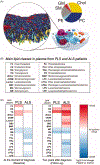
Pathological changes in the main classes of lipids in plasma from PLS and ALS patients. A. Computer simulation of the spatial distribution of lipids and cholesterol in the outer leaflet of the plasma membrane. Shown are 63 different lipid species, combining 14 types of headgroups and 11 types of tails. Cholesterols are colored yellow, lipid headgroups are colored by type: Phosphatidylcholine PC blue, Sphingomyelin SM gray, Phospatidylethanolamine PE cyan; GM red; Phosphatidylinositolphosphates PIP magenta; Phosphatidylinositol PI pink; Phosphatidylserin PS green; PA, white; CE, ice blue; Diacylglycerol brown; LPC, orange. The pie chart shows the relative distribution of the main lipid in the outer leaflet of the plasma membrane. Modified from (84) with the kind permission of the authors and the publisher. B. Table showing the main classes of lipids in plasma from PLS and ALS patients analyzed by LC-MS. C. Heat map representation of the most significant fold-changes in the concentration of every class of lipids in plasma from ALS and PLS patients compared to controls at the beginning of the study (baseline) and two years after (Follow-up). (n = 40 samples analyzed in triplicate. *<0.05; **<0.01. t-test).

Similar articles

Cited by

References

    1. Erb WH. Ueber einen wenig bekanten spinalen Symptomenkomplex. About a little-known spinal symptom complex. Berliner Klinische Wochenschrift 1875;26:357–9.
    1. Stark FM, Moersch FP. Primary lateral sclerosis. A distinct clinical entity. J Nerv Mental Dis. 1945;102:332–7.
    1. Pringle CE, Hudson AJ, Munoz DG, Kiernan JA, Brown WF, Ebers GC. Primary lateral sclerosis. Clinical features, neuropathology and diagnostic criteria. Brain 1992;115: 495–520. - PubMed
    1. Gordon PH, Cheng B, Katz IB, Pinto M, Hays AP, Mitsumoto H, et al. The natural history of primary lateral sclerosis. Neurology 2006;66:647–53. - PubMed
    1. Mitsumoto H, Nagy PL, Gennings C, Murphy J, Andrews H, Goetz R, et al. Phenotypic and molecular analyses of primary lateral sclerosis. Neurol Genet. 2015;1:e3. - PMC - PubMed

Publication types

Substances