

Morphological and genetic heterogeneity of synchronous multifocal lung adenocarcinoma in a Chinese cohort
- PMID: 33602172
- PMCID: PMC7890910
- DOI: 10.1186/s12885-021-07892-8
Morphological and genetic heterogeneity of synchronous multifocal lung adenocarcinoma in a Chinese cohort
Abstract
Background: Synchronous multifocal lung cancer (SMLC) is diagnosed with increasing frequency in clinical practice globally. Due to innate variation in clinical management and outcome, it is vital to properly distinguish between synchronous multifocal primary lung cancer (SMPLC) and intrapulmonary metastasis (IM). The pathologic features and principal classification criteria of multifocal lung cancer remain unclear. Our objective was to evaluate the diagnostic value of histological morphologic features and driver gene mutations in SMLC classification.
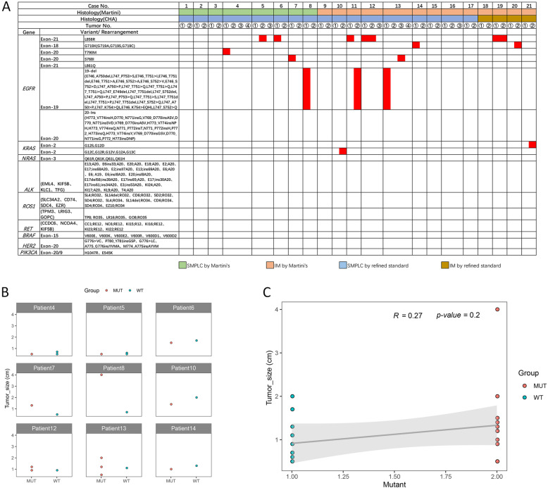
Methods: We collected a unique cohort of Chinese patients with SMLC, and fully explored the morphologic, immunohistochemical, and molecular features of the disease. Twenty-one SMLC patients with a total of 50 tumours were included in our study. The pathological features that were presented by these patients were analysed, including the tumours location, tumours size, pathological types, predominant pattern of adenocarcinoma, and immunohistochemical staining. We conducted molecular testing of nine driver oncogenes that are associated with lung cancer, namely, EGER, KRAS, BRAF, NRAS, ALK, ROS1, RET, HER2, and PIK3CA.
Results: According to the Martini-Melamed classification and refined standard, 8 and 17 patients, respectively, were considered to have SMPLCs. Gene mutations were identified in 18 tumours (36%). Twelve patients had different gene mutations.
Conclusions: We demonstrate that conventional morphological assessment is not sufficient to clearly establish the clonal relationship of SMPLCs. Instead, the evaluation of histological subtypes, including nonmucinous adherent components, is required. Multiplex genotypic analysis may also prove to be a useful additional tool.
Keywords: Morphological assessment; Multiplex genotypic analysis; Synchronous multifocal lung cancer (SMLC); Synchronous multifocal primary lung cancer (SMPLC).
Conflict of interest statement
The authors declare that they have no competing interests.
Figures


Similar articles
-
Molecular profiling of key driver genes improves staging accuracy in multifocal non-small cell lung cancer.J Thorac Cardiovasc Surg. 2020 Aug;160(2):e71-e79. doi: 10.1016/j.jtcvs.2019.11.126. Epub 2019 Dec 20. J Thorac Cardiovasc Surg. 2020. PMID: 32007245
-
Concomitant EGFR Mutation and EML4-ALK Rearrangement in Lung Adenocarcinoma Is More Frequent in Multifocal Lesions.Clin Lung Cancer. 2019 Jul;20(4):e517-e530. doi: 10.1016/j.cllc.2019.04.008. Epub 2019 Apr 25. Clin Lung Cancer. 2019. PMID: 31138506
-
Molecular diagnostic characteristics based on the next generation sequencing in lung cancer and its relationship with the expression of PD-L1.Pathol Res Pract. 2020 Feb;216(2):152797. doi: 10.1016/j.prp.2019.152797. Epub 2019 Dec 23. Pathol Res Pract. 2020. PMID: 31926773
-
Concordant and Discordant EGFR Mutations in Patients With Multifocal Adenocarcinomas: Implications for EGFR-Targeted Therapy.Clin Ther. 2016 Jul;38(7):1567-76. doi: 10.1016/j.clinthera.2016.06.005. Epub 2016 Jun 29. Clin Ther. 2016. PMID: 27368115 Free PMC article. Review.
-
Surgical Prognosis of Synchronous Multiple Primary Lung Cancer: Systematic Review and Meta-Analysis.Clin Lung Cancer. 2021 Jul;22(4):341-350.e3. doi: 10.1016/j.cllc.2020.10.022. Epub 2020 Nov 5. Clin Lung Cancer. 2021. PMID: 33243621
Cited by
-
Differential Diagnostic Value of Histology in MPLC and IPM: A Systematic Review and Meta-Analysis.Front Oncol. 2022 Apr 29;12:871827. doi: 10.3389/fonc.2022.871827. eCollection 2022. Front Oncol. 2022. PMID: 35574321 Free PMC article.
-
Intertumoural Heterogeneity and Branch Evolution of Synchronous Multiple Primary Lung Adenocarcinomas by Next-Generation Sequencing Analysis.Front Oncol. 2021 Nov 3;11:760715. doi: 10.3389/fonc.2021.760715. eCollection 2021. Front Oncol. 2021. PMID: 34804960 Free PMC article.
-
Diagnosis and management of multiple primary lung cancer.Front Oncol. 2024 Oct 1;14:1392969. doi: 10.3389/fonc.2024.1392969. eCollection 2024. Front Oncol. 2024. PMID: 39411141 Free PMC article. Review.
-
Case Report: Response to ALK-TKIs in a metastatic lung cancer patient with morphological heterogeneity and consistent molecular features.Front Oncol. 2023 Aug 11;13:1209799. doi: 10.3389/fonc.2023.1209799. eCollection 2023. Front Oncol. 2023. PMID: 37637057 Free PMC article.
References
-
- Detterbeck FC, Franklin WA, Nicholson AG, et al. The IASLC lung cancer staging project: background data and proposed criteria to distinguish separate primary lung cancers from metastatic foci in patients with two lung tumors in the forthcoming eighth edition of the TNM classification for lung cancer. J Thorac Oncol. 2016;11(5):651–665. doi: 10.1016/j.jtho.2016.01.025. - DOI - PubMed
-
- Travis WD, Brambilla E, Burke AP, Marx A, Nicholson AG, editors. WHO classification of tumors of the lung, pleura, thymus and heart. 4. Lyon: France IARC Press; 2015. - PubMed
MeSH terms
Grants and funding
LinkOut - more resources
Full Text Sources
Other Literature Sources
Medical
Research Materials
Miscellaneous
