. 2021 Feb 18;18(1):46.
doi: 10.1186/s12974-021-02095-1.

Platelets and lymphocytes drive progressive penumbral tissue loss during middle cerebral artery occlusion in mice

Affiliations

Platelets and lymphocytes drive progressive penumbral tissue loss during middle cerebral artery occlusion in mice

Michael K Schuhmann et al. J Neuroinflammation. .

Abstract

Background: In acute ischemic stroke, cessation of blood flow causes immediate tissue necrosis within the center of the ischemic brain region accompanied by functional failure in the surrounding brain tissue designated the penumbra. The penumbra can be salvaged by timely thrombolysis/thrombectomy, the only available acute stroke treatment to date, but is progressively destroyed by the expansion of infarction. The underlying mechanisms of progressive infarction are not fully understood.

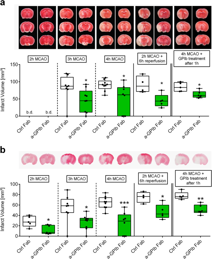
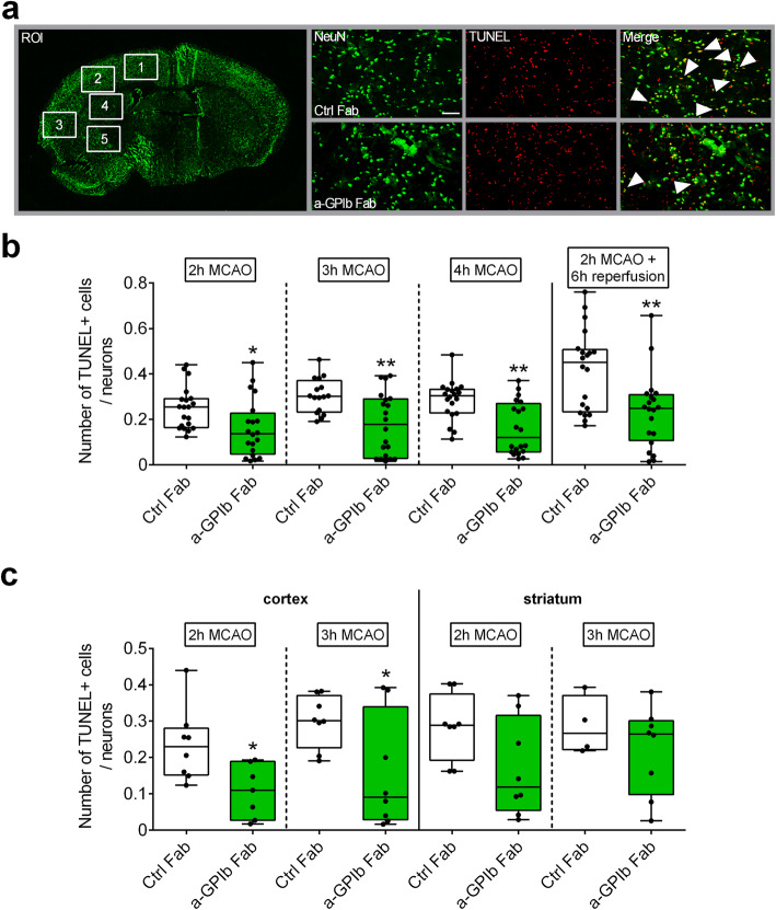
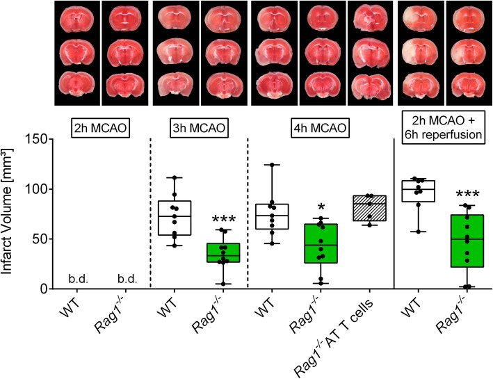
Methods: To address mechanisms, mice underwent filament occlusion of the middle cerebral artery (MCAO) for up to 4 h. Infarct development was compared between mice treated with antigen-binding fragments (Fab) against the platelet surface molecules GPIb (p0p/B Fab) or rat immunoglobulin G (IgG) Fab as control treatment. Moreover, Rag1-/- mice lacking T-cells underwent the same procedures. Infarct volumes as well as the local inflammatory response were determined during vessel occlusion.

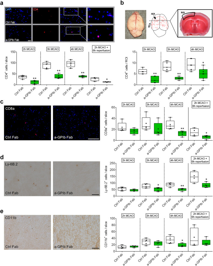
Results: We show that blocking of the platelet adhesion receptor, glycoprotein (GP) Ibα in mice, delays cerebral infarct progression already during occlusion and thus before recanalization/reperfusion. This therapeutic effect was accompanied by decreased T-cell infiltration, particularly at the infarct border zone, which during occlusion is supplied by collateral blood flow. Accordingly, mice lacking T-cells were likewise protected from infarct progression under occlusion.

Conclusions: Progressive brain infarction can be delayed by blocking detrimental lymphocyte/platelet responses already during occlusion paving the way for ultra-early treatment strategies in hyper-acute stroke before recanalization.

Keywords: Glycoprotein receptor Ib; Ischemic penumbra; Ischemic stroke; T-cells; Thrombo-inflammation; middle cerebral artery occlusion.


Conflict of interest statement

The authors declare no competing interests.

Figures

Fig. 1
Fig. 1
Blocking of GPIb delays ischemic brain damage. Representative images of coronal sections stained with a TTC, b Map2a/b 2, 3 and 4 h after MCAO or after 2 h of MCAO with additional 6 h of reperfusion in mice treated with rat IgG Fab (Ctrl Fab) or p0p/B Fab (a-GPIb Fab) immediately or 1 h after MCA occlusion. Infarcted areas are shown in white. Planimetric analyses were used to quantify the infarct volumes. Results are presented as box plots (n = 4–9). *P< 0.05, **P< 0.01, ***P< 0.001 between the indicated groups, 2-tailed Student t test or, in the case of nonparametric functional outcome, the Wilcoxon–Mann–Whitney U-test was applied. b.d., beyond detection level
Fig. 2
Fig. 2
Neuronal apoptosis is reduced with GPIb treatment. a Schematic view of brain regions of interest (ROI) analyzed to quantify TUNEL+ cells/neurons and representative immunocytologic stainings. b Quantification of neurons stained with NeuN (Alexa488, green) and subjected to TUNEL assay (TMR red, red) in the ipsilateral hemisphere 2, 3, and 4 h after MCAO or after 2 h of MCAO with additional 6 h of reperfusion overall (ROI 1-5) and c divided in cortex (ROI 1 and 2: neocortex) and striatum (ROI 4 and 5: subcortex) stained with NeuN (Alexa488, green) and subjected to TUNEL assay (TMR red, red) in the ipsilateral hemisphere 2 and 3 h after MCAO in mice treated with rat IgG Fab (Ctrl Fab) or p0p/B Fab (a-GPIb Fab) using × 20 objective. Scale bar 100 μm (n = 16–20 slides). *P< 0.05, **P< 0.01 between the indicated groups, 2-tailed Student t test or, in the case of nonparametric functional outcome, the Wilcoxon–Mann–Whitney U-test was applied
Fig. 3
Fig. 3
Blocking of platelet GPIb diminished ultra-early T-cell recruitment in the ischemic brain. a Representative immunocytologic stainings (top) and quantification (bottom) of brain-infiltrating CD4-positive T lymphocytes (Cy3, red), nuclei (DAPI, blue) in the whole ipsilateral hemisphere 2, 3, and 4 h after MCAO or after 2 h of MCAO with additional 6 h of reperfusion in mice treated with rat IgG Fab (Ctrl Fab) or p0p/B Fab (a-GPIb Fab) using × 20 objective. Scale bar 100 μm (n = 4–5). b (top) Native whole mouse brain with schematic overview of ACA and MCA territories. The ROI used for T-cell quantification is shown on a TTC-stained coronal brain section (2 h MCAO). (bottom) Quantification of brain-infiltrating CD4-positive T lymphocytes (Cy3, red), nuclei (DAPI, blue) in the ROI at 2, 3, and 4 h after MCAO in mice treated with rat IgG Fab (Ctrl Fab) or p0p/B Fab (a-GPIb Fab) using × 20 objective (n = 5). Representative images (left) and quantification (right) of c CD8a-positive cells (CD8a (Alexa 647, red), nuclei (DAPI, blue), d Ly-6B.2-positive cells, or e CD11b-positive cells (3,3-Diaminobenzidin (DAB)) in the ipsilateral hemisphere 2, 3, and 4 h after MCAO or after 2 h of MCAO with additional 6 h of reperfusion in mice treated with rat IgG Fab (Ctrl Fab) or p0p/B Fab (a-GPIb Fab) using × 20 objective. Scale bar 100 μm (n = 4–5). *P< 0.05, **P< 0.01 between the indicated groups, 2-tailed Student t test or, in the case of nonparametric functional outcome, the Wilcoxon–Mann–Whitney U-test was applied. ACA, anterior cerebral artery; MCA, middle cerebral artery; ROI, region of interest
Fig. 4
Fig. 4
T-cells are required to promote infarct growth during MCAO. Representative images of coronal sections stained with TTC 2, 3, and 4 h after MCAO or after 2 h of MCAO with additional 6 h of reperfusion in Rag1−/− or WT mice and representative images of coronal sections stained with TTC 4 h after MCAO in Rag1−/− mice with adoptively transferred T-cells (AT T-cells). Infarcted areas are shown in white. Planimetric analysis was used to quantify the infarct volume. Results are presented as box plots (n = 5–10). *P< 0.05, and ***P< 0.001 between the indicated groups, 2-tailed Student t test or, in the case of nonparametric functional outcome, the Wilcoxon–Mann–Whitney U-test was applied. Nonparametric functional outcome between 3 groups were compared by Kruskal–Wallis test with post hoc Dunn’s multiple comparisons test. b.d., beyond detection level

References

    1. Goyal M, Menon BK, van Zwam WH, Dippel DW, Mitchell PJ, Demchuk AM, Davalos A, Majoie CB, van der Lugt A, de Miquel MA, et al. Endovascular thrombectomy after large-vessel ischaemic stroke: a meta-analysis of individual patient data from five randomised trials. Lancet. 2016;387:1723–1731. doi: 10.1016/S0140-6736(16)00163-X. - DOI - PubMed
    1. Stoll G, Nieswandt B. Thrombo-inflammation in acute ischaemic stroke - implications for treatment. Nat Rev Neurol. 2019;15:473–481. doi: 10.1038/s41582-019-0221-1. - DOI - PubMed
    1. Mizuma A, You JS, Yenari MA. Targeting reperfusion injury in the age of mechanical thrombectomy. Stroke. 2018;49:1796–1802. doi: 10.1161/STROKEAHA.117.017286. - DOI - PMC - PubMed
    1. Nour M, Scalzo F, Liebeskind DS. Ischemia-reperfusion injury in stroke. Interv Neurol. 2013;1:185–199. doi: 10.1159/000353125. - DOI - PMC - PubMed
    1. Eltzschig HK, Eckle T. Ischemia and reperfusion--from mechanism to translation. Nat Med. 2011;17:1391–1401. doi: 10.1038/nm.2507. - DOI - PMC - PubMed

Substances

LinkOut - more resources