. 2021 Feb 18;18(1):50.
doi: 10.1186/s12974-021-02092-4.

Critical roles of sphingosine kinase 1 in the regulation of neuroinflammation and neuronal injury after spinal cord injury

Affiliations

Critical roles of sphingosine kinase 1 in the regulation of neuroinflammation and neuronal injury after spinal cord injury

Chenjian Wang et al. J Neuroinflammation. .

Abstract

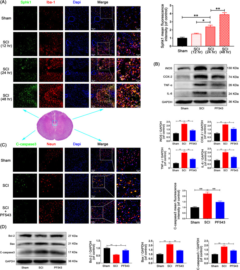
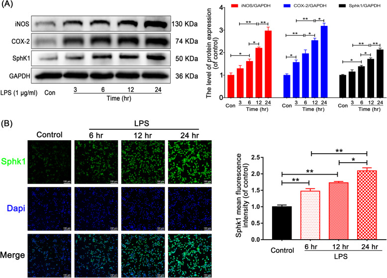
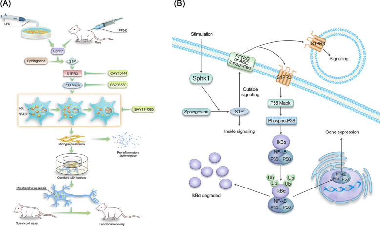
Background: The pathological process of traumatic spinal cord injury (SCI) involves excessive activation of microglia leading to the overproduction of proinflammatory cytokines and causing neuronal injury. Sphingosine kinase 1 (Sphk1), a key enzyme responsible for phosphorylating sphingosine into sphingosine-1-phosphate (S1P), plays an important role in mediating inflammation, cell proliferation, survival, and immunity.

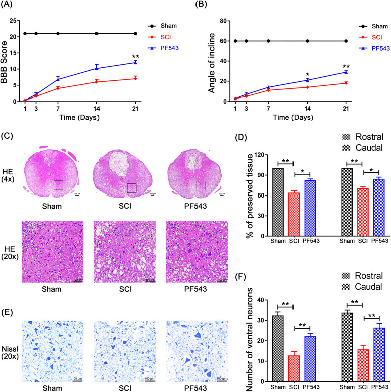
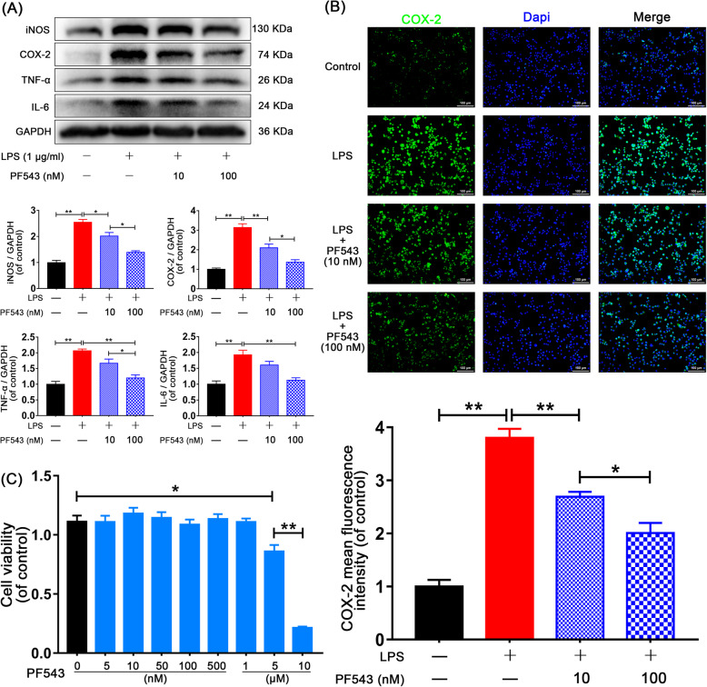
Methods: We aim to investigate the mechanism and pathway of the Sphk1-mediated neuroinflammatory response in a rodent model of SCI. Sixty Sprague-Dawley rats were randomly assigned to sham surgery, SCI, or PF543 (a specific Sphk1 inhibitor) groups. Functional outcomes included blinded hindlimb locomotor rating and inclined plane test.

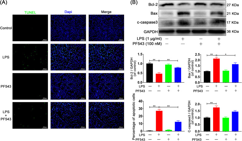
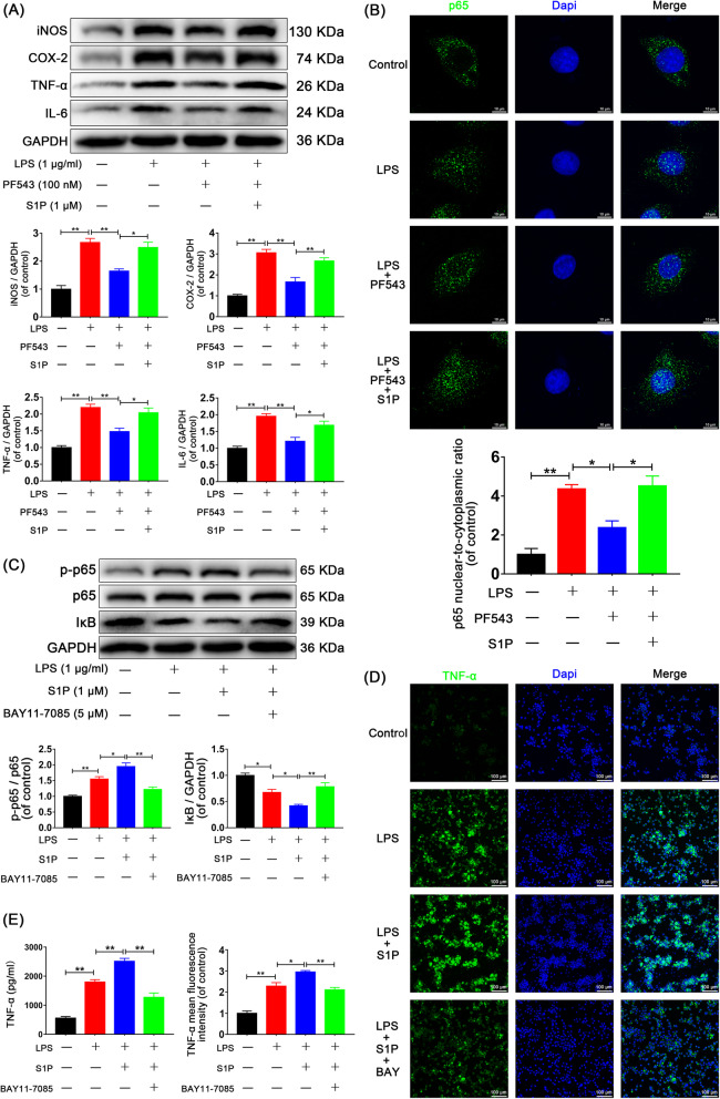
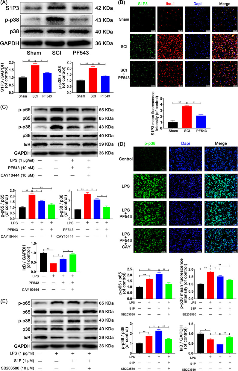
Results: We discovered that Sphk1 is upregulated in injured spinal cord tissue of rats after SCI and is associated with production of S1P and subsequent NF-κB p65 activation. PF543 attenuated p65 activation, reduced inflammatory response, and relieved neuronal damage, leading to improved functional recovery. Western blot analysis confirmed that expression of S1P receptor 3 (S1PR3) and phosphorylation of p38 mitogen-activated protein kinase (p38 MAPK) are activated in microglia of SCI rats and mitigated by PF543. In vitro, we demonstrated that Bay11-7085 suppressed NF-κB p65 and inhibited amplification of the inflammation cascade by S1P, reducing the release of proinflammatory TNF-α. We further confirmed that phosphorylation of p38 MAPK and activation of NF-κB p65 is inhibited by PF543 and CAY10444. p38 MAPK phosphorylation and NF-κB p65 activation were enhanced by exogenous S1P and inhibited by the specific inhibitor SB204580, ultimately indicating that the S1P/S1PR3/p38 MAPK pathway contributes to the NF-κB p65 inflammatory response.

Conclusion: Our results demonstrate a critical role of Sphk1 in the post-traumatic SCI inflammatory cascade and present the Sphk1/S1P/S1PR3 axis as a potential target for therapeutic intervention to control neuroinflammation, relieve neuronal damage, and improve functional outcomes in SCI.

Keywords: M1 microglia; NF-κB p65 activation; S1P/S1PR3/p38 MAPK pathway; Sphk1; Spinal cord injury.


Conflict of interest statement

The authors declare that they have no competing interests.

Figures

Fig. 1
Fig. 1
Inhibition of Sphk1 attenuates microglial M1 polarization and prevents neuronal apoptosis in SCI rats. a Double-staining for Sphk1 (green)/Iba-1 (red) in injured spinal cord tissue from each group of rats (scale bar: 100 μm or 50 μm; 5 mm caudal from injury core). Sphk1 was induced over time in activated microglia of SCI rats. b Western blot analysis of iNOS, COX-2, TNF-α, IL-6, and GAPDH expression in each group of rats. M1 polarization and inflammatory mediator release was suppressed by PF543 treatment. c Representative Western blots of cleaved caspase 3, Bcl-2, Bax, and GAPDH expression in each group of rats. PF543 prevented SCI-induced neuronal apoptosis. d Double-staining for cleaved caspase 3 (green)/NeuN (red) in sections of injured spinal cord tissue from each group of rats (scale bar: 100 μm or 50 μm). n = 3 per group. *P < 0.05 and **P < 0.01
Fig. 2
Fig. 2
PF543 reduces structural tissue damage, reduces neuron loss, and promotes the recovery of motor function after experimental acute spinal cord injury. a Basso, Beattie and Bresnahan (BBB) scores. b Inclined plane test scores. c H&E stains at 7 days. Scale bars are 200 μm (× 4) and 100 μm (× 20). Dashed line area shows cavity of spinal cord. d Graphic presentation of the percentage of preserved tissue relative to the transverse area of the spinal cord on the seventh day after surgery. e Nissl staining to assess the loss of neurons at 7 days (scale bar: 100 μm). f Counting analysis of ventral neurons at rostral 5 mm and caudal 5 mm. n = 5 per group. *P < 0.05 and **P < 0.01
Fig. 3
Fig. 3
LPS induces M1 microglia polarization and Sphk1 expression in HAPI cells. a Representative Western blots and quantitative data for iNOS, COX-2, and Sphk1, GAPDH expression in each group of HAPI cells. LPS induced M1 microglia polarization and Sphk1 expression in a time-dependent manner. b Immunofluorescence staining for Sphk1 in each group of HAPI cells (scale bar: 100 μm). n = 3 independent experiments. *P < 0.05 and **P < 0.01
Fig. 4
Fig. 4
PF543 attenuates neuroinflammation in vitro. HAPI cells were pretreated with 10 nM and 100 nM PF543 for 12 h, followed by LPS treatment for 24 h. a Representative Western blots and quantitative data for iNOS, COX-2, TNF-a, IL-6, and GAPDH expression in each group of microglia. PF543 suppressed proinflammatory mediator release and prevented M1 microglia polarization in a concentration-dependent manner. b Immunofluorescence staining for COX-2 in each of group microglia (scale bar: 100 μm). c HAPI cells were treated with different concentrations of PF543 for 48 h and then the cell viability was evaluated by CCK-8 assay. n = 3 independent experiments. *P < 0.05 and **P < 0.01
Fig. 5
Fig. 5
PF543 prevents M1 microglia-induced neuronal apoptosis. a TUNEL assay was performed in neurons cocultured with microglia (scale bar: 100 μm). The neuronal apoptosis induced by M1 microglia was significantly attenuated in cells cocultured with PF543 pretreated microglia. b Representative Western blots and quantitative data for Bcl-2, Bax, cleaved caspase 3, and GAPDH expression in each group of neurons. n = 3 independent experiments. *P < 0.05 and **P < 0.01
Fig. 6
Fig. 6
Sphk1-regulated neuroinflammation is related to S1P-induced NF-κB activation. HAPI cells were pretreated with PF543 for 12 h or BAY11-7085 for 2 h, followed by washing three times and treatment with LPS alone or LPS plus S1P for 24 h. a Representative Western blots and quantitative data for iNOS, COX-2, TNF-α, IL-6, and GAPDH expression. PF543 pretreatment attenuated LPS-induced M1 microglia polarization and proinflammatory cytokine production, effects that were reversed by addition of exogenous S1P. b Immunofluorescence staining for p65 was observed under confocal microscope (scale bar: 10 μm), and nuclear-to-cytoplasmic ratio of p65 was quantified. LPS-induced p65 nuclear translocation was suppressed by PF543 pretreatment and restored by addition of exogenous S1P. c Western blot analysis of p-p65, p65, IκB, and GAPDH expression in HAPI cells. Adding exogenous S1P amplified the NF-κB activation and inflammatory response, and BAY11-7085 significantly eliminated the amplification. d Immunofluorescence staining for TNF-α in each group of microglia (scale bar: 100 μm). e The secretion of TNF-α was also measured by ELISA assay. S1P promoted proinflammatory mediator release, and the effect was eliminated by BAY11-7085. n = 3 independent experiments. *P < 0.05 and **P < 0.01
Fig. 7
Fig. 7
S1P/S1PR3/p38 pathway contributes to Sphk1-related p65 activation. For in vivo experiments, rats were sacrificed and specimens were taken at 3 days after SCI. n = 3 per group. a Representative Western blots and quantitative data for S1PR3, p-p38, p38, and GAPDH expression in each group of rats. S1PR3 expression and p38 phosphorylation were enhanced in the SCI group and were inhibited by PF543 treatment. b Double-staining for S1PR3 (green)/Iba-1 (red) in injured spinal cord tissue from each group of rats (scale bar: 100 μm). For in vitro studies, HAPI cells were pretreated with PF543 for 12 h and CAY10444 for 2 h or SB203580 for 2 h, followed by washing three times and treatment with LPS alone or LPS plus S1P for 24 h, n = 3 independent experiments. c Representative Western blots and quantitative data for p-p65, p65, p-p38, p38, IκB, and GAPDH expression in each group of microglia. Both Sphk1 and S1PR3 activity influenced the activation of p65 and p38. d Immunofluorescence staining for p-p38 in each of group microglia (scale bar: 100 μm), PF543 pretreatment decreased p38 phosphorylation, and inhibition of S1PR3 strengthened the inhibitory effect. e Western blot analysis of p-p65, p65, p-p38, p38, IκB, and GAPDH expression in each group of microglia. S1P enhanced the activation of p65 and p38 induced by LPS stimulation, the effect was reversed by SB203580. *P < 0.05 and **P < 0.01
Fig. 8
Fig. 8
a Schematic of the S1P/S1PR3/p38 MAPK signaling pathway involved in neuroinflammation. After LPS stimulation or spinal cord injury, resting microglia underwent M1 polarization and released proinflammatory mediators to induce neuronal apoptosis, which was improved by the specific inhibitors. b One of the Sphk1/S1P signaling pathways. S1P catalyzed by Sphk1 in the cytoplasm are transported outside the cell and bind to membrane receptors (S1PR3), then downstream activation of p38 MAPK phosphorylation and NF-κB nuclear transcription is triggered

References

    1. Spinal cord injury (SCI) 2016 facts and figures at a glance. J Spinal Cord Med. 2016;39:493–494. doi: 10.1080/10790268.2016.1210925. - DOI - PMC - PubMed
    1. Ramer L, Ramer M, Bradbury E. Restoring function after spinal cord injury: towards clinical translation of experimental strategies. Lancet Neurol. 2014;13:1241–1256. doi: 10.1016/S1474-4422(14)70144-9. - DOI - PubMed
    1. Cannon B. Sensation and loss. Nature. 2013;503:S2–S3. doi: 10.1038/503S2a. - DOI - PubMed
    1. Hu X, Leak R, Shi Y, Suenaga J, Gao Y, Zheng P, Chen J. Microglial and macrophage polarization—new prospects for brain repair. Nat Rev Neurol. 2015;11:56–64. doi: 10.1038/nrneurol.2014.207. - DOI - PMC - PubMed
    1. Cherry J, Olschowka J, O'Banion M. Neuroinflammation and M2 microglia: the good, the bad, and the inflamed. J Neuroinflam. 2014;11:98. doi: 10.1186/1742-2094-11-98. - DOI - PMC - PubMed

MeSH terms

LinkOut - more resources