. 2021 May 1;27(9):2613-2623.
doi: 10.1158/1078-0432.CCR-20-4436. Epub 2021 Feb 18.

Clinicopathologic and Genomic Analysis of TP53-Mutated Endometrial Carcinomas

Affiliations

Clinicopathologic and Genomic Analysis of TP53-Mutated Endometrial Carcinomas

Amir Momeni-Boroujeni et al. Clin Cancer Res. .

Abstract

Purpose: Copy number-high endometrial carcinomas were described by The Cancer Genome Atlas as high-grade endometrioid and serous cancers showing frequent copy-number alterations (CNA), low mutational burden (i.e., non-hypermutant), near-universal TP53 mutation, and unfavorable clinical outcomes. We sought to investigate and compare the clinicopathologic and molecular characteristics of non-hypermutant TP53-altered endometrial carcinomas of four histologic types.

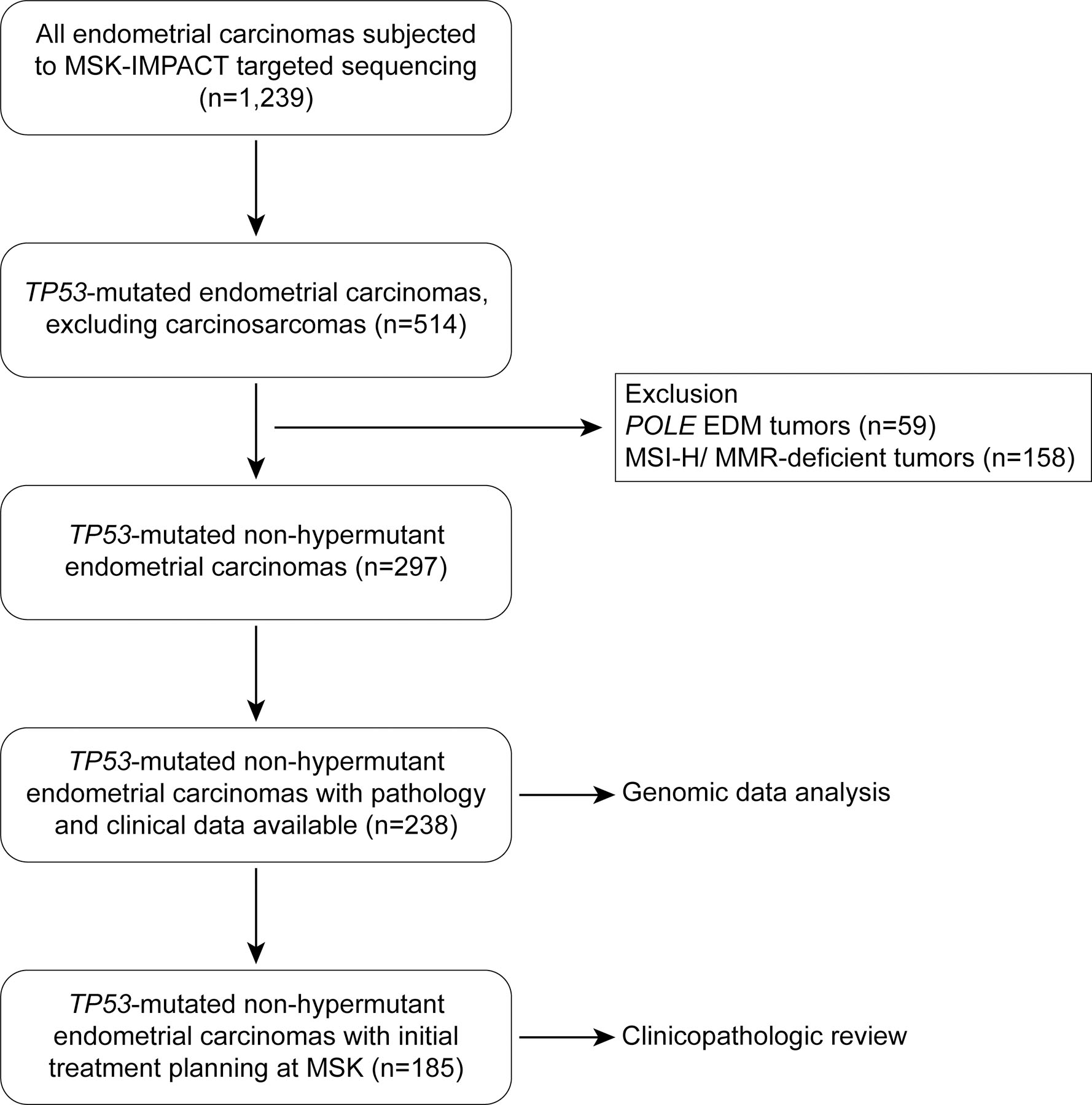
Experimental design: TP53-mutated endometrial carcinomas, defined as TP53-mutant tumors lacking microsatellite instability or pathogenic POLE mutations, were identified (n = 238) in a cohort of 1,239 endometrial carcinomas subjected to clinical massively parallel sequencing of 410-468 cancer-related genes. Somatic mutations and CNAs (n = 238), and clinicopathologic features were determined (n = 185, initial treatment planning at our institution).

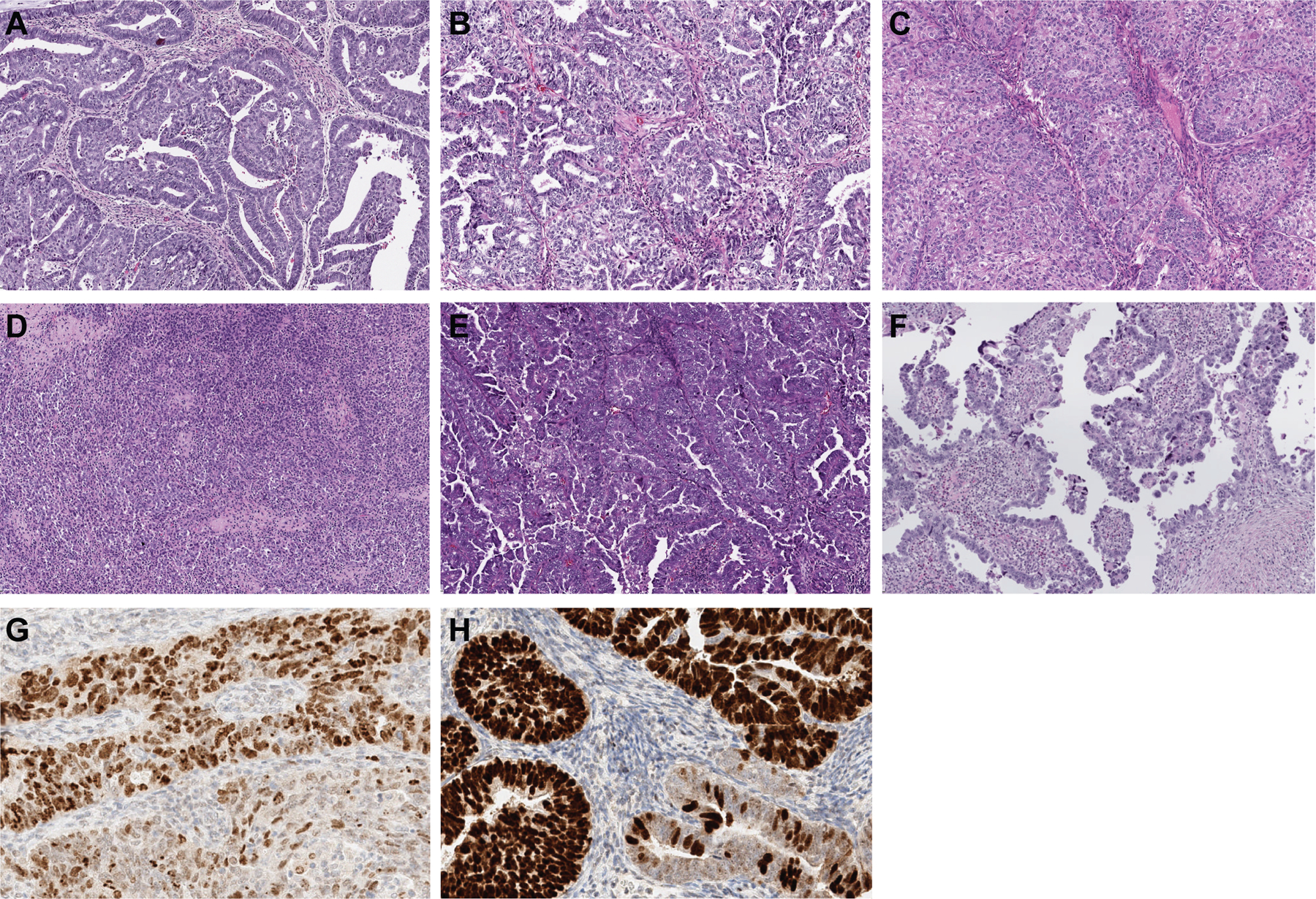
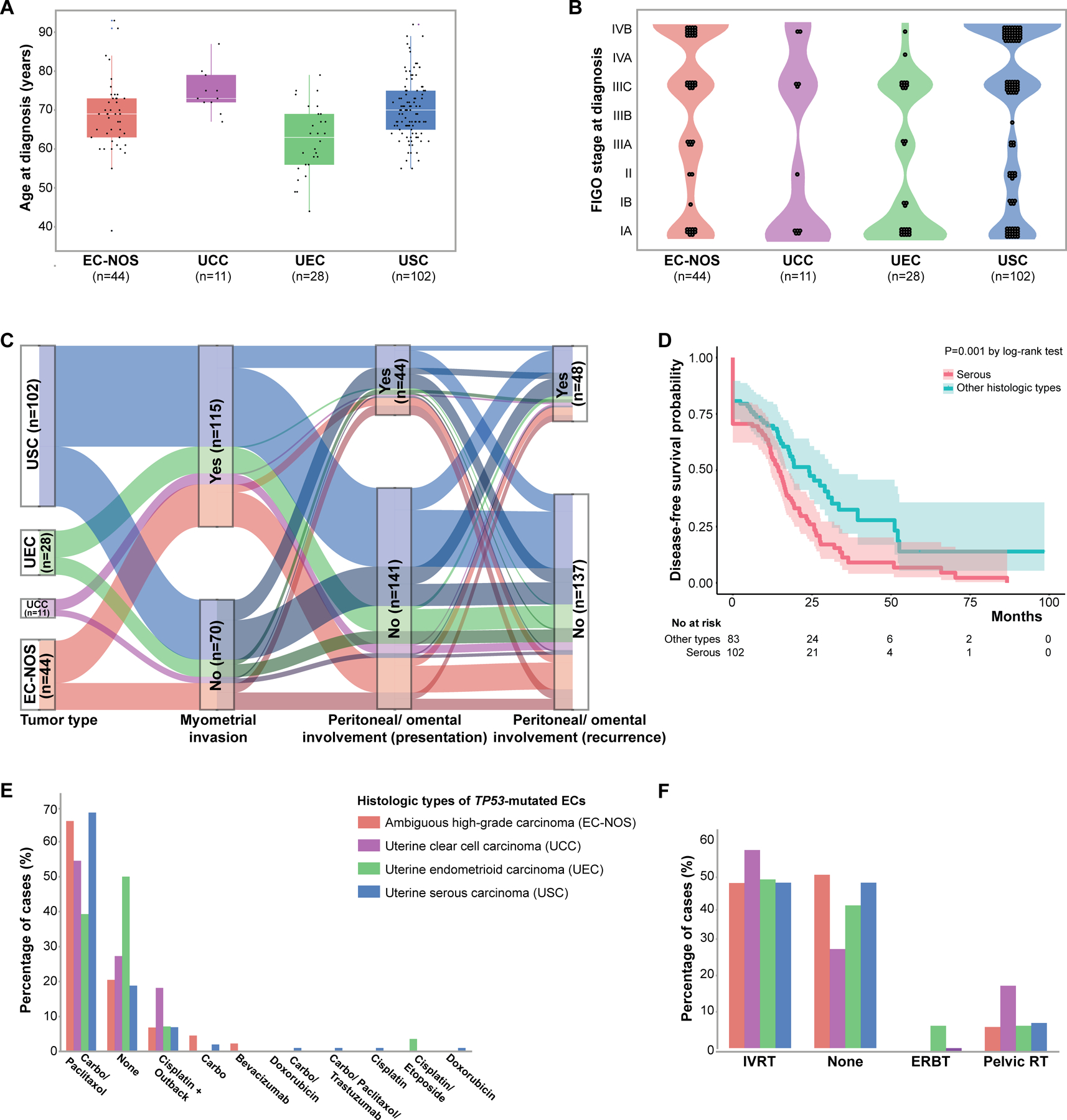
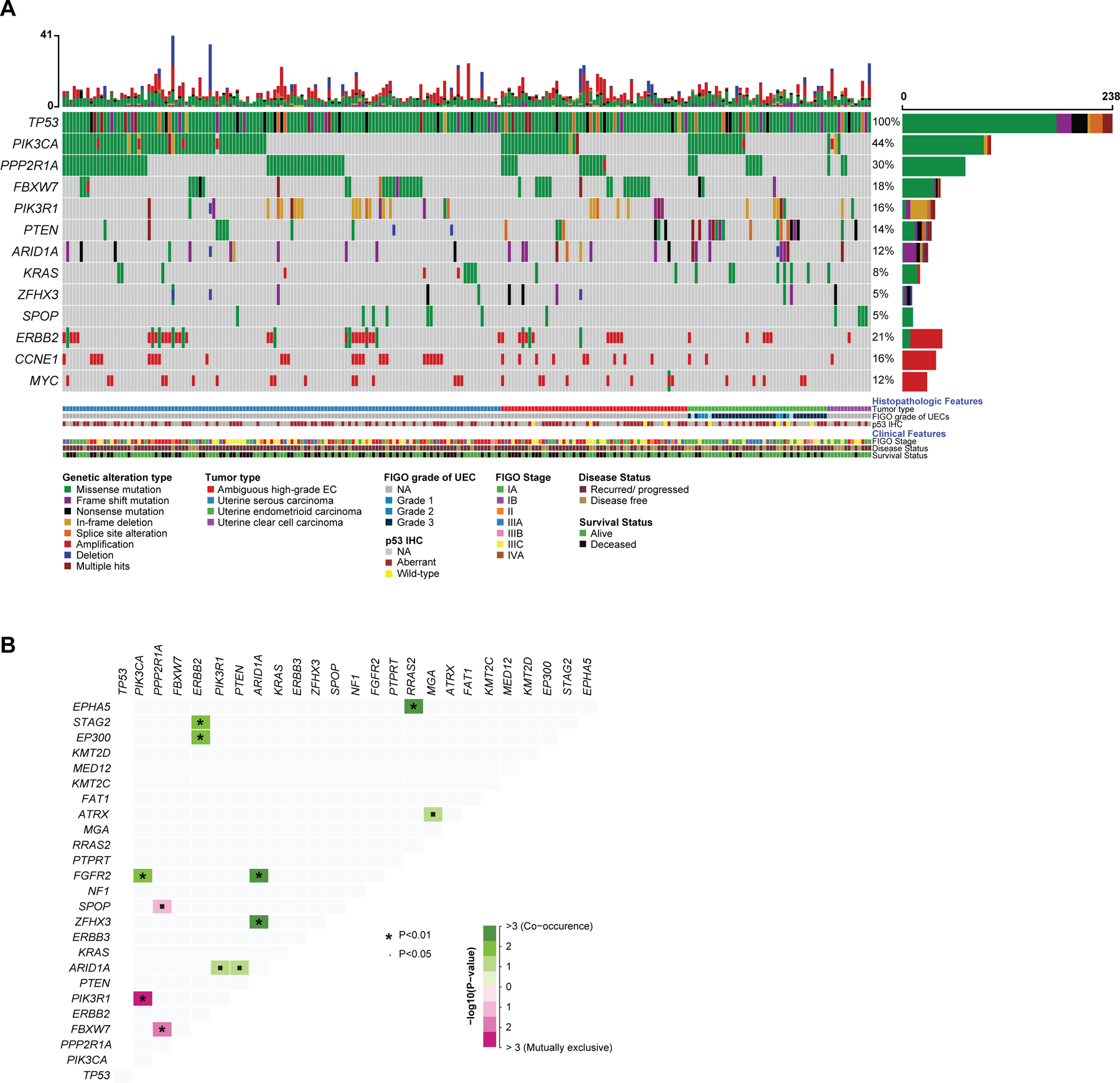
Results: TP53-mutated endometrial carcinomas encompassed uterine serous (n = 102, 55.1%), high-grade endometrial carcinoma with ambiguous features/not otherwise specified (EC-NOS; n = 44, 23.8%), endometrioid carcinomas of all tumor grades (n = 28, 15.1%), and clear cell carcinomas (n = 11, 5.9%). PTEN mutations were significantly more frequent in endometrioid carcinomas, SPOP mutations in clear cell carcinomas, and CCNE1 amplification in serous carcinomas/EC-NOS; however, none of these genomic alterations were exclusive to any given histologic type. ERBB2 amplification was present at similar frequencies across TP53-mutated histologic types (7.7%-18.6%). Although overall survival was similar across histologic types, serous carcinomas presented more frequently at stage IV, had more persistent and/or recurrent disease, and reduced disease-free survival.

Conclusions: TP53-mutated endometrial carcinomas display clinical and molecular similarities across histologic subtypes. Our data provide evidence to suggest performance of ERBB2 assessment in all TP53-mutated endometrial carcinomas. Given the distinct clinical features of serous carcinomas, histologic classification continues to be relevant.


Conflict of interest statement

Conflicts of interest: None declared.

Figures

Figure 1.
Figure 1.. CONSORT diagram summarizing the selection process of non-hypermutant TP53-mutated endometrial cancers included in this study
EDM, exonuclease domain mutant; MMR, DNA mismatch repair; MSI, microsatellite instability
Figure 2.
Figure 2.. Histologic subtypes of non-hypermutant TP53-mutated endometrial carcinomas and p53 expression
TP53-mutated endometrial carcinomas are of different histologic subtypes. TP53-mutated endometrioid endometrial carcinoma can present as (A) FIGO grade 1, (B) grade 2, or (C) grade 3. (D) TP53-mutated endometrial carcinomas can have ambiguous morphologic features. (E) The most common histologic subtype is endometrial serous carcinoma. (F) Endometrial clear cell carcinoma is the least common morphologic variant. In some TP53-mutated endometrial carcinomas, p53 aberrant expression can be subclonal. The subclonal aberrant expression in (G) shows variable overexpression of p53 in most cells of UEC (top), whereas the subclonal aberrant expression in (H), a high-grade endometrial carcinoma with ambiguous features/ not otherwise specified (EC-NOS), shows more uniform overexpression of p53 (top), the bottom glands in both (G) and (H) show wild-type (heterogenous) p53 staining pattern.
Figure 3.
Figure 3.. Clinicopathologic features of non-hypermutant TP53-mutated endometrial carcinomas and association with outcome
(A) Age at TP53-mutated endometrial cancer diagnosis. These tumors mostly occur in elderly patients. (B) FIGO stage at TP53-mutated endometrial cancer diagnosis. The majority of TP53-mutated tumors present with advanced FIGO stages. (C) Alluvial graph showing myometrial invasion, peritoneal/ omental involvement at presentation and recurrence in patients with TP53-mutated endometrial carcinomas. Note that TP53-mutated endometrial cancers have similar rates of peritoneal involvement irrespective of histologic subtype. Cases with non-myoinvasive disease are shown in darker shades. (D), Kaplan-Meier curve for disease-free survival of patients with TP53-mutated endometrial cancer of serous histology and those of other histologic subtypes (i.e. clear cell, endometrioid, and ambiguous high-grade). (E) Percentage of patients with TP53-mutated endometrial cancer who received adjuvant chemotherapy. Note that the choice of adjuvant therapy was different between UEC and USC. (Carbo: carboplatin). (F) Percentage of patients with TP53-mutated endometrial cancer who received adjuvant radiation therapy. IVRT, intravascular radiation therapy; ERBT, External beam radiation therapy; RT, radiotherapy. EC-NOS, high-grade endometrial carcinoma with ambiguous features/ not otherwise specified; UCC, uterine clear cell carcinoma; UEC, uterine endometrioid carcinoma; USC, uterine serous carcinoma.
Figure 4.
Figure 4.. Somatic mutations and gene copy number alterations in non-hypermutant TP53-mutated endometrial carcinomas
(A) Oncoprint depicting the most recurrent genomic alterations in TP53-mutated endometrial carcinomas. Each column represents a tumor with the bar graph at the top depicting the number/distribution of alterations per sample, and the Oncoprint rows showing alterations for each gene. The bottom part of the graph shows the summary of histopathologic and clinical information for each case. The bar graph on the right of the panel shows the number and distribution of alterations for each gene. Mutation types and clinicopathologic features are color-coded according to the legend. NA, not available. (B) Distance matrix showing the somatic interaction of commonly altered genes in TP53-mutated endometrial carcinomas. Significant co-occurrences are marked in shades of green and the significant mutual exclusivity marked in shades of purple. The size of the * depicting the p-value and the intensity of color shows the degree of association or exclusion.

References

    1. Cancer Genome Atlas Research Network, Kandoth C, Schultz N, Cherniack AD, Akbani R, Liu Y, et al. Integrated genomic characterization of endometrial carcinoma. Nature 2013;497:67–73. - PMC - PubMed
    1. Kommoss S, McConechy MK, Kommoss F, Leung S, Bunz A, Magrill J, et al. Final validation of the ProMisE molecular classifier for endometrial carcinoma in a large population-based case series. Ann Oncol 2018;29:1180–8. - PubMed
    1. DeLair DF, Burke KA, Selenica P, Lim RS, Scott SN, Middha S, et al. The genetic landscape of endometrial clear cell carcinomas. J Pathol 2017;243:230–41. - PMC - PubMed
    1. Cherniack AD, Shen H, Walter V, Stewart C, Murray BA, Bowlby R, et al. Integrated Molecular Characterization of Uterine Carcinosarcoma. Cancer Cell 2017;31:411–23. - PMC - PubMed
    1. Kim SR, Cloutier BT, Leung S, Cochrane D, Britton H, Pina A, et al. Molecular subtypes of clear cell carcinoma of the endometrium: Opportunities for prognostic and predictive stratification. Gynecol Oncol 2020;158:3–11. - PubMed

Publication types

MeSH terms