. 2021 Feb 19;371(6531):846-849.
doi: 10.1126/science.abe0763.

Mechanism of membrane-tethered mitochondrial protein synthesis

Affiliations

Mechanism of membrane-tethered mitochondrial protein synthesis

Yuzuru Itoh et al. Science. .

Abstract

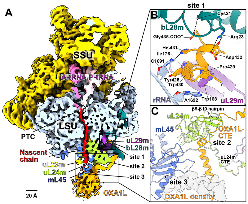
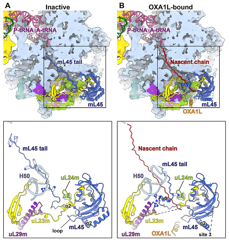
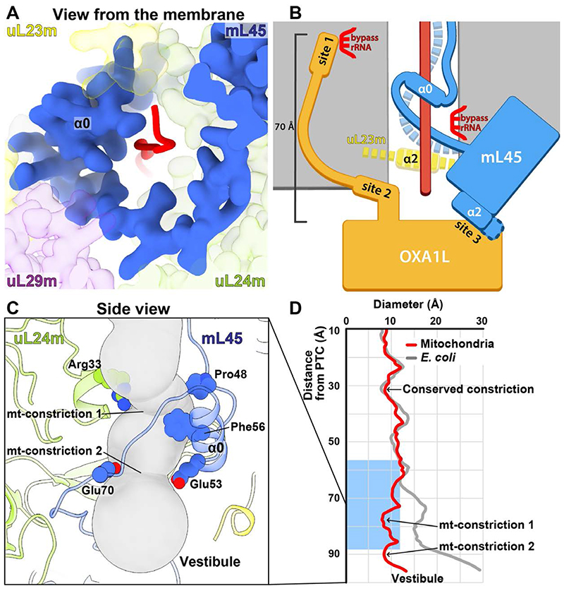
Mitochondrial ribosomes (mitoribosomes) are tethered to the mitochondrial inner membrane to facilitate the cotranslational membrane insertion of the synthesized proteins. We report cryo-electron microscopy structures of human mitoribosomes with nascent polypeptide, bound to the insertase oxidase assembly 1-like (OXA1L) through three distinct contact sites. OXA1L binding is correlated with a series of conformational changes in the mitoribosomal large subunit that catalyze the delivery of newly synthesized polypeptides. The mechanism relies on the folding of mL45 inside the exit tunnel, forming two specific constriction sites that would limit helix formation of the nascent chain. A gap is formed between the exit and the membrane, making the newly synthesized proteins accessible. Our data elucidate the basis by which mitoribosomes interact with the OXA1L insertase to couple protein synthesis and membrane delivery.


Conflict of interest statement

Competing interests: Authors declare no competing interests.

Figures

Fig. 1
Fig. 1. Human mitoribosome:OXAlL complex.
(A) Density of mitoribosome:OXA1L complex lowpass-filtered to 6 Å resolution for clarity and cut through the tunnel. Contact sites 1, 2, 3 are indicated. (B) Contact site 1 is formed by OXA1L C-terminal helix bound to bL28m, uL29m, and rRNA. Tyr428 is stacked on rRNA C1691 and His431 hydrogen-bonds with C1691. The backbone carbonyl and sidechain of Asp432 form polar interactions with bL28m (Arg23, Pro429, and Trp430) and hydrophobic interactions with uL29m (Trp168, Ile176), and rRNA A1692. (C) The OXA1L-CTE interacts with uL24m (site 2), and the OXA1L core (shown with density) is associated with mL45-α2 (site 3).
Fig. 2
Fig. 2. Biochemical characterization of mL45 and OXA1L interaction.
Immunoprecipitation of mL45 from mitochondrial extracts of T-Rex-293 cells expressing wild-type (WT) or ΔN-mL45, and the DDX28-KO cells overexpressing WT mL45. The cells were probed for mL45, OXA1L, or bL12m as an mtLSU marker, IgG was used as a control. (IP) immunoprecipitated, (UB) unbound, (T) total input.
Fig. 3
Fig. 3. Comparison between inactive and OXA1L-bound structures.
(A) In the inactive state, the mL45 N-terminal tail occupies the tunnel. Basic residues in the tunnel are shown as spheres in the bottom panel. uL23m-α2 is stabilized by the mL45 tail and a loop (residues 200-208) of mL45. The tip of H50 is disordered due to flexibility. (B) In the OXA1L-bound state, a nascent polypeptide occupies the tunnel. The mL45 tail is folded and stabilized by uL24m, uL29m, and rRNA H50. The basic residues participating in the stabilization are shown in the bottom panel. The tip of H50 is ordered, uL23m-α2, and the mL45 loop are disordered, and the path through the tunnel is open. The contact site 3 with OXA1L is formed via mL45-α2. The bottom panels show partial structures of uL23m (residues 37-153), uL24m (93-112), and uL29m (94-157).
Fig. 4
Fig. 4. Mechanism of membrane-tethered nascent polypeptide emergence.
(A) Folding of the mL45 tail stabilized by uL29m and uL24m, resulting in a continuous protein arch coating the interior with the nascent chain. (B) Sideview schematic of the gating mechanism. The folding of mL45-α0 (blue tube) leads to the detachment of its N-terminus from rRNA (dashed blue) and destabilization of uL23m-α2 (dashed yellow) that together open the way for a polypeptide chain (red). OXA1L is docked to the extended mL45-α2 (site 3) for co-translational insertion of the emerging polypeptide. Contact sites 1 and 2 are formed between the OXA1L-CTE and the mitoribosomal surface up to 70 Å away from the membrane surface. (C) Sideview of the lower tunnel, shown as a gray tube. Mitochondria-specific constriction sites are formed by mL45-α0 and uL24m, and the involved residues are shown. (D) The tunnel diameter was calculated for the mitoribosome and E. coli ribosome and plotted along the path. The blue rectangle indicates the position of an α-helical formation within the tunnel in bacteria.

References

    1. De Silva D, Tu Y-T, Amunts A, Fontanesi F, Barrientos A. Mitochondrial ribosome assembly in health and disease. Cell Cycle. 2015;14:2226–2250. - PMC - PubMed
    1. Suomalainen A, Battersby BJ. Mitochondrial diseases: the contribution of organelle stress responses to pathology. Nat Rev Mol Cell Biol. 2018;19:77–92. - PubMed
    1. Aibara S, Singh V, Modelska A, Amunts A. Structural basis of mitochondrial translation. Elife. 2020;9:1–17. - PMC - PubMed
    1. Englmeier R, Pfeffer S, Förster F. Structure of the Human Mitochondrial Ribosome Studied In Situ by Cryoelectron Tomography. Structure. 2017;25:1574–1581. e2. - PubMed
    1. Chen Y, Capponi S, Zhu L, Gellenbeck P, Freites JA, White SH, Dalbey RE. YidC Insertase of Escherichia coli: Water Accessibility and Membrane Shaping. Structure. 2017;25:1403–1414. e3. - PMC - PubMed

Publication types

MeSH terms